Finding a 5mm lung nodule on your chest imaging can understandably cause significant anxiety. These small pulmonary lesions, roughly the size of a peppercorn, represent one of the most common incidental findings in modern medical imaging. The overwhelming majority of 5mm nodules are benign , with studies consistently showing that less than 1% prove to be malignant. However, the discovery of any pulmonary nodule requires careful evaluation and appropriate surveillance to ensure optimal patient outcomes. Understanding the classification systems, risk stratification models, and evidence-based management protocols can help demystify the clinical approach to these small but potentially significant findings.
Understanding 5mm pulmonary nodule classification and detection methods
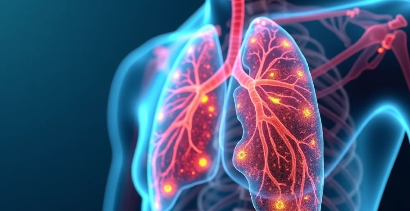
The detection and characterisation of 5mm pulmonary nodules relies heavily on advanced imaging techniques and standardised classification systems. These small lesions often represent the lower threshold of detection capabilities, making accurate assessment both challenging and critically important for patient care.
High-resolution computed tomography (HRCT) imaging protocols for small nodules
Modern HRCT protocols have revolutionised the detection of small pulmonary nodules, with slice thicknesses of 1.25mm or less enabling precise visualisation of lesions as small as 2-3mm. The technical parameters for optimal 5mm nodule detection include low radiation dose protocols, typically employing 120kVp tube voltage and modulated tube current based on patient body habitus. Contemporary CT scanners can achieve excellent image quality whilst maintaining radiation exposure below 1.5 mSv per examination , making serial surveillance both safe and practical.
Reconstruction algorithms play a crucial role in nodule detection accuracy. Iterative reconstruction techniques, such as adaptive statistical iterative reconstruction (ASIR) or model-based iterative reconstruction (MBIR), significantly improve image quality whilst reducing noise. These advances have increased the sensitivity for detecting 5mm nodules from approximately 85% with conventional techniques to over 95% with current protocols.
Radiological appearance characteristics: Ground-Glass vs solid nodule morphology
The radiological appearance of 5mm nodules provides essential information for risk stratification and management decisions. Solid nodules appear as homogeneous, well-defined lesions with density similar to soft tissue structures. These lesions completely obscure underlying pulmonary vessels and parenchyma, creating a sharp interface with surrounding lung tissue.
Ground-glass nodules present as hazy areas of increased attenuation that do not obscure underlying bronchial and vascular markings. Part-solid nodules combine elements of both morphologies, containing areas of ground-glass opacity with focal solid components. Interestingly, pure ground-glass nodules at 5mm demonstrate the lowest malignancy rates , often representing inflammatory changes or atypical adenomatous hyperplasia rather than invasive carcinoma.
Fleischner society guidelines for 5mm nodule management
The Fleischner Society guidelines provide evidence-based recommendations for managing small pulmonary nodules, including specific protocols for 5mm lesions. For solid nodules measuring exactly 5mm, the guidelines distinguish between low-risk and high-risk patient populations. Low-risk patients, typically defined as non-smokers under 60 years without significant occupational exposures, may not require routine follow-up imaging for 5mm solid nodules.
High-risk patients, including current or former smokers with significant pack-year histories, require more intensive surveillance. The guidelines recommend optional CT follow-up at 12 months for 5mm solid nodules in high-risk individuals. For ground-glass nodules measuring 5mm, no routine follow-up is generally recommended regardless of risk factors, reflecting their extremely low malignancy potential.
Low-dose CT screening programme identification rates
Large-scale lung cancer screening programmes have provided extensive data on 5mm nodule prevalence and outcomes. The National Lung Screening Trial (NLST) reported that approximately 15% of screening participants had nodules measuring 4-6mm on baseline examination. Among these small nodules, the cancer detection rate was remarkably low, at less than 0.5% for 5mm lesions.
European screening programmes, including the NELSON trial, have reported similar findings with slightly different size thresholds. The volume-based approach used in NELSON classified nodules measuring 4.2-10mm as intermediate risk , with follow-up protocols adjusted accordingly. These programmes demonstrate that whilst 5mm nodules are relatively common in screening populations, the vast majority remain stable or resolve spontaneously during surveillance periods.
Risk stratification using established clinical prediction models
Accurate risk assessment for 5mm pulmonary nodules requires sophisticated prediction models that incorporate multiple clinical and radiological variables. These validated tools help clinicians make informed decisions about surveillance intensity and intervention timing whilst minimising unnecessary anxiety and healthcare costs.
Brock university nodule risk calculator application
The Brock University prediction model, also known as the PanCan model, represents one of the most widely validated tools for assessing malignancy probability in small pulmonary nodules. This model incorporates eight variables: patient age, sex, smoking history, family history of lung cancer, emphysema presence, nodule location, nodule type, and nodule diameter. For 5mm nodules, the model typically generates probability estimates ranging from 1-5% in most clinical scenarios.
The Brock model demonstrates particular strength in identifying very low-risk nodules that may safely avoid intensive surveillance. When the calculated probability falls below 5%, the model reliably identifies nodules with minimal malignancy risk , supporting less aggressive management approaches. Online calculators make this tool readily accessible to clinicians worldwide, improving standardisation of risk assessment practices.
Mayo clinic nodule risk model assessment parameters
The Mayo Clinic model, whilst historically important, shows limitations when applied specifically to 5mm nodules. This model incorporates six variables: age, smoking history, cancer history, nodule diameter, upper lobe location, and spiculation. However, the model was derived from a population with larger nodules, potentially overestimating cancer probability in very small lesions.
Recent validation studies suggest that the Mayo Clinic model may generate probability estimates 2-3 times higher than actual cancer rates for 5mm nodules. This limitation has led many institutions to favour more contemporary models, such as the Brock University calculator, which incorporate larger datasets and more diverse patient populations.
Herder prediction model for indeterminate pulmonary nodules
The Herder model, developed from a Dutch screening programme, provides another validated approach to 5mm nodule risk assessment. This model incorporates demographic factors, nodule characteristics, and CT appearance features to generate probability estimates. The Herder model demonstrates particular accuracy for nodules in the 5-10mm size range, making it especially relevant for borderline lesions.
One distinctive feature of the Herder model is its incorporation of nodule texture analysis and edge characteristics. For 5mm nodules with smooth, well-defined borders, the model typically generates probability estimates below 2% , supporting conservative management approaches. The model has undergone extensive external validation across multiple European populations, demonstrating consistent performance across different healthcare systems.
Pancan model integration in clinical Decision-Making
The PanCan model has emerged as the preferred tool for many thoracic specialists managing 5mm nodules. Its development from the largest available dataset, including over 12,000 nodules from multiple international centres, provides robust statistical foundations. The model’s online interface allows real-time probability calculations during clinical encounters, facilitating shared decision-making with patients.
Integration of PanCan model results into clinical practice requires careful consideration of confidence intervals and model limitations. For 5mm nodules, the model performs optimally when probability estimates fall into clearly low-risk (<5%) or intermediate-risk (5-15%) categories. The model’s accuracy decreases for nodules with unusual characteristics or in patients with atypical demographic profiles , necessitating clinical judgement in these scenarios.
Differential diagnosis: benign vs malignant 5mm lesions
Distinguishing benign from malignant causes of 5mm pulmonary nodules requires systematic evaluation of patient history, imaging characteristics, and epidemiological factors. The differential diagnosis encompasses infectious, inflammatory, and neoplastic aetiologies, each with distinct clinical presentations and imaging features.
Infectious causes represent the most common aetiology for 5mm pulmonary nodules, particularly in younger patients and those with recent respiratory symptoms. Granulomatous infections, including histoplasmosis, coccidioidomycosis, and tuberculosis, frequently manifest as small, well-defined nodules that may calcify over time. Geographic distribution plays a crucial role in infectious differential diagnosis , with histoplasmosis predominating in the Ohio and Mississippi river valleys, whilst coccidioidomycosis occurs primarily in southwestern United States regions.
Inflammatory nodules secondary to rheumatoid arthritis, Wegener’s granulomatosis, or sarcoidosis typically appear as multiple bilateral lesions with upper lobe predominance. These nodules often demonstrate associated mediastinal lymphadenopathy or other systemic manifestations that aid in diagnosis. Hamartomas, representing the most common benign neoplasm, characteristically display popcorn calcification patterns on CT imaging, though this feature may not be apparent in 5mm lesions.
Primary lung carcinomas measuring 5mm are exceedingly rare but theoretically possible. Adenocarcinoma represents the most likely histological subtype at this size, often presenting as part-solid lesions with ground-glass components. Squamous cell carcinomas rarely manifest as 5mm nodules, typically requiring larger dimensions before becoming radiologically apparent. Metastatic disease from extrapulmonary primary tumours, whilst possible, uncommonly presents as isolated 5mm lesions except in cases of haematogenous dissemination from renal cell carcinoma or thyroid carcinoma.
Statistical analysis from major screening programmes demonstrates that malignancy probability for 5mm nodules remains consistently below 1% across all demographic groups, providing reassurance for both patients and clinicians managing these common findings.
Evidence-based surveillance protocols and Follow-Up imaging
Surveillance strategies for 5mm pulmonary nodules must balance the need for early cancer detection against the risks of overdiagnosis and patient anxiety. Evidence-based protocols have evolved significantly following large-scale screening trials and longitudinal observational studies that provide robust outcome data for small nodule management.
British thoracic society recommendations for small nodule monitoring
The British Thoracic Society (BTS) guidelines offer a risk-stratified approach to 5mm nodule surveillance that considers both patient factors and nodule characteristics. For solid nodules measuring 5-6mm in low-risk patients, the BTS recommends no routine follow-up, acknowledging the extremely low malignancy probability. High-risk patients with 5mm solid nodules warrant consideration of 12-month follow-up CT scanning, though this remains optional rather than mandatory.
The BTS guidelines emphasise the importance of patient counselling and shared decision-making in surveillance planning. For anxious patients who prefer active monitoring despite low risk, compromise surveillance protocols may be appropriate , typically involving single follow-up examination at 12 months. The guidelines also stress the importance of smoking cessation counselling for all patients with pulmonary nodules, regardless of size or risk stratification.
Interval imaging timeframes: 3-month vs 12-month CT protocols
The optimal timing for follow-up imaging of 5mm nodules remains a subject of ongoing research and clinical debate. Traditional approaches favoured shorter intervals, with 3-month follow-up examinations designed to detect rapid growth suggestive of malignancy. However, contemporary evidence suggests that such intensive surveillance may be unnecessary for most 5mm lesions.
Recent analysis of screening programme data demonstrates that clinically significant growth in 5mm nodules occurs rarely within the first year of detection. Mathematical modelling studies indicate that extending surveillance intervals to 12 months for low-risk 5mm nodules results in negligible delays in cancer diagnosis whilst substantially reducing radiation exposure and healthcare costs. This evidence supports current guideline recommendations for longer initial surveillance intervals.
Volumetric growth rate assessment using Computer-Aided detection
Volumetric analysis represents a significant advancement in 5mm nodule surveillance, offering more precise growth detection than traditional diameter-based measurements. Computer-aided detection (CAD) systems can calculate nodule volumes with submillimetre precision, enabling detection of growth rates as low as 25% volume increases. This sensitivity proves particularly valuable for 5mm nodules where small absolute changes represent significant proportional increases.
Volume doubling time calculations provide quantitative metrics for distinguishing benign from malignant growth patterns. Benign nodules typically demonstrate volume doubling times exceeding 400 days, whilst malignant lesions show doubling times between 100-300 days. For 5mm nodules showing volume increases exceeding 25% at first follow-up, additional evaluation may be warranted regardless of absolute size criteria . CAD systems also improve inter-observer reproducibility, reducing measurement variability that historically complicated small nodule surveillance.
When to discharge from radiological surveillance programmes
Determining appropriate endpoints for 5mm nodule surveillance requires consideration of stability duration, patient risk factors, and resource allocation. Current evidence supports surveillance termination after two years of stability for most 5mm nodules, based on data showing extremely low malignancy rates among nodules remaining unchanged over this timeframe.
The decision to discharge patients from surveillance programmes should incorporate individual risk assessment and patient preferences. High-risk patients with multiple nodules or strong family histories may warrant extended surveillance beyond standard timeframes. Conversely, young non-smokers with isolated 5mm ground-glass nodules may safely discontinue surveillance after shorter periods. Clear communication about discharge decisions helps patients understand that surveillance termination reflects low cancer probability rather than inadequate medical care .
Advanced diagnostic interventions for persistent 5mm nodules
The vast majority of 5mm pulmonary nodules require only surveillance rather than invasive diagnostic procedures. However, specific clinical scenarios may warrant consideration of advanced diagnostic interventions, particularly when nodules demonstrate growth, develop concerning morphological features, or occur in extremely high-risk patients with strong clinical suspicion of malignancy.
Positron emission tomography (PET) scanning demonstrates limited utility for 5mm nodules due to resolution limitations and high false-negative rates. Most PET scanners cannot reliably detect metabolic activity in lesions smaller than 8-10mm, making this modality inappropriate for routine 5mm nodule evaluation. False-negative PET results occur in up to 50% of malignant nodules measuring 5-8mm , potentially providing false reassurance and delaying appropriate treatment.
Percutaneous biopsy techniques face significant technical challenges when targeting 5mm lesions. CT-guided needle biopsy requires precise localisation and carries substantial pneumothorax risk, particularly for peripheral lesions. The small target size increases sampling error rates and may necessitate multiple attempts, escalating complication risks. Most interventional radiologists prefer larger targets (>8mm) for percutaneous procedures, though highly experienced operators may successfully sample 5mm lesions in selected cases.
Advanced bronchoscopic techniques, including electromagnetic navigation and robotic-assisted procedures, have expanded diagnostic capabilities for small peripheral nodules, though success rates remain suboptimal for 5mm lesions located beyond segmental bronchi.
Surgical intervention for 5mm nodules remains controversial and typically reserved for exceptional circumstances. Video-assisted thoracoscopic surgery (VATS) wedge resection can successfully remove 5mm lesions whilst preserving maximal lung function. However, the morbidity and mortality associated with surgical intervention rarely justify proceeding based solely on a 5mm nodule, even in high-risk patients. Most thoracic surgeons prefer to observe 5mm nodules until they reach 8-10mm before considering surgical intervention , unless multiple concerning features coexist.
Patient counselling: communicating lung cancer risk and anxiety management
Effective patient communication about 5mm pulmonary nodules requires balancing honest risk assessment with appropriate reassurance. Many patients experience significant anxiety following nodule detection, often overestimating cancer probability and catastrophising potential outcomes. Healthcare providers must address these concerns whilst maintaining vigilance for the small subset of nodules requiring intervention.
Risk communication should employ clear, accessible language that avoids medical jargon whilst maintaining accuracy. Explaining
that 5mm nodules represent less than a 1% chance of cancer helps patients understand their actual risk level. Visual aids, such as icon arrays showing 99 green circles and 1 red circle, can effectively illustrate these low probability statistics. Comparative risk frameworks, such as explaining that the chance of cancer in a 5mm nodule is similar to the risk of being struck by lightning in a given year, provide relatable context for patients struggling with abstract probability concepts.
Healthcare providers should acknowledge patient anxiety whilst reinforcing the evidence-based rationale for surveillance approaches. Discussing the harms of overdiagnosis and unnecessary procedures helps patients understand why immediate biopsy or surgical intervention is typically not recommended for 5mm nodules. Encouraging questions and providing written materials for home review supports informed decision-making and reduces post-consultation anxiety.
Practical anxiety management strategies should complement medical information. Recommending stress reduction techniques, such as mindfulness meditation or deep breathing exercises, can help patients cope with surveillance periods. Establishing clear follow-up schedules and providing direct contact information for questions between appointments reduces uncertainty and improves patient satisfaction with surveillance protocols.
Patient education should emphasise controllable risk factors, particularly smoking cessation for current smokers. Discussing the substantial impact of continued smoking on lung cancer risk provides motivation for behaviour change whilst empowering patients to actively reduce their cancer probability. For former smokers, reinforcing the benefits of smoking cessation and discouraging relapse supports long-term health outcomes beyond nodule management.
The psychological impact of 5mm nodule detection often exceeds the actual medical significance, making effective communication and anxiety management essential components of comprehensive patient care.
Regular reassessment of patient understanding and emotional well-being throughout surveillance periods ensures ongoing support. Some patients may benefit from referral to specialist lung cancer screening coordinators or clinical nurse specialists with expertise in nodule management. These professionals can provide additional counselling time and follow-up support that busy clinical schedules may not accommodate. Documentation of patient preferences regarding surveillance intensity and communication frequency facilitates continuity of care across multiple providers and institutions.
Shared decision-making frameworks should guide surveillance planning, particularly for borderline cases where guidelines offer flexible recommendations. Involving patients in risk-benefit discussions about surveillance frequency, radiation exposure, and anxiety management creates collaborative care plans that align with individual values and preferences. This approach improves adherence to surveillance protocols whilst respecting patient autonomy in healthcare decisions affecting their quality of life.