Discovering black residue on a cotton swab after cleaning your ears can be an unsettling experience that prompts immediate concern about your ear health. This dark material may range from deep brown to jet black and can appear as flakes, sticky deposits, or powdery substances. While the sight of black earwax might initially alarm you, understanding the various underlying causes can help distinguish between benign conditions and those requiring medical attention. The colour and consistency of ear discharge provide valuable diagnostic clues for healthcare professionals, as different pathological processes produce distinctly different appearances in ceruminous secretions.
The external auditory canal serves as a complex ecosystem where multiple factors influence the production, composition, and colour of earwax. Various microorganisms, environmental contaminants, and physiological processes can alter the normal appearance of cerumen, sometimes resulting in dramatically darkened secretions. Recognising the potential causes behind black ear residue enables you to make informed decisions about when to seek professional medical evaluation versus managing the condition with conservative measures.
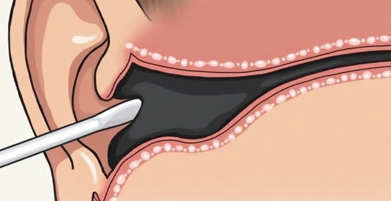
Cerumen impaction and excessive earwax production
Cerumen impaction represents one of the most common causes of darkened ear discharge, affecting millions of individuals worldwide. When earwax accumulates over extended periods, oxidation processes naturally darken the cerumen from its typical yellow or amber colour to deep brown or black. The transformation occurs gradually as lipids within the wax undergo chemical changes similar to how cooking oils darken when exposed to heat and air over time.
The impaction process typically develops when cerumen production exceeds the ear’s natural self-cleaning mechanisms. Normal jaw movements during chewing, talking, and yawning help migrate earwax from deep within the canal toward the outer opening. However, when this natural conveyor belt system becomes disrupted, wax begins accumulating in layers, eventually hardening and darkening through dehydration and oxidative processes.
Sebaceous gland hyperactivity in the external auditory canal
Sebaceous glands within the ear canal can become hyperactive due to hormonal fluctuations, stress, or genetic predisposition, leading to excessive wax production. This overproduction creates an environment where cerumen cannot exit the ear efficiently, resulting in progressive accumulation. The increased sebaceous activity often correlates with similar conditions affecting facial skin, such as acne or seborrhoeic dermatitis, suggesting systemic rather than localised glandular dysfunction.
Hormonal influences during puberty, pregnancy, or menopause frequently exacerbate sebaceous gland activity throughout the body, including those within the auditory canal. Additionally, certain medications, particularly those affecting hormone levels or skin oil production, can indirectly influence cerumen consistency and production rates, creating favourable conditions for impaction development.
Keratin accumulation from desquamated epithelial cells
The ear canal’s epithelial lining continuously sheds dead skin cells as part of normal tissue renewal processes. These desquamated cells, rich in keratin protein, become incorporated into cerumen and contribute significantly to its bulk and colour. When excessive epithelial shedding occurs due to inflammatory conditions, allergic reactions, or mechanical trauma, the increased keratin content can dramatically darken the resulting wax mixture.
Chronic scratching or aggressive cleaning with cotton swabs often triggers increased epithelial turnover, creating a cycle where mechanical irritation leads to more cell shedding, which in turn contributes to greater wax accumulation. The keratin-rich debris tends to form cohesive masses that resist natural migration, further promoting impaction and colour changes.
Wet-type vs Dry-Type cerumen genetic polymorphisms
Genetic variations in the ABCC11 gene determine whether individuals produce wet or dry cerumen, with significant implications for impaction risk and colour development. Wet-type cerumen, more common in individuals of African and European descent, contains higher lipid concentrations that make it stickier and more prone to accumulation. This consistency change increases the likelihood of impaction and subsequent darkening through oxidative processes.
Dry-type cerumen, predominantly found in East Asian populations, typically appears lighter and flakier but can still undergo colour changes when impacted. The genetic polymorphism affects not only wax consistency but also the activity levels of ceruminous glands, influencing overall production volumes and the propensity for accumulation-related colour changes.
Age-related changes in ceruminous gland function
Advancing age brings predictable changes to ceruminous gland function, with older adults experiencing increased wax viscosity and reduced natural clearance mechanisms. The aging process affects both the quantity and quality of cerumen production, often resulting in drier, more adhesive wax that resists normal migration patterns. These age-related changes significantly increase impaction risk and the likelihood of observing darkened ear discharge.
Elderly individuals also experience decreased jaw mobility and reduced skin elasticity within the ear canal, further compromising natural wax clearance mechanisms. The combination of altered wax composition and reduced clearance creates ideal conditions for extensive accumulation and the dramatic colour changes associated with chronic impaction.
Otomycosis and fungal infections in the ear canal
Fungal infections of the external ear canal frequently produce distinctive black or very dark brown discharge that can easily be mistaken for impacted cerumen. Otomycosis affects approximately 7% of individuals seeking treatment for ear-related symptoms, with certain fungal species producing characteristic pigmented secretions. The warm, humid environment of the ear canal provides ideal growing conditions for various fungi, particularly in individuals who frequently expose their ears to moisture through swimming or humid climates.
Fungal overgrowth typically develops when the ear’s natural protective mechanisms become compromised through excessive cleaning, minor trauma, or underlying skin conditions. The resulting infection produces inflammatory exudates that mix with fungal spores and metabolic byproducts, creating the dark, often malodorous discharge characteristic of otomycosis. Unlike simple cerumen impaction, fungal infections usually accompany symptoms such as itching, pain, and a sensation of fullness within the affected ear.
Aspergillus niger spore colonisation patterns
Aspergillus niger represents one of the most distinctive fungal pathogens affecting the ear canal, producing characteristically black spores that create dramatic dark discharge. This opportunistic organism thrives in warm, moist environments and can rapidly colonise compromised ear canal tissue. The black pigmentation results from melanin production within the fungal spores, creating discharge that appears almost coal-black when examined on cotton swabs.
The colonisation pattern of Aspergillus niger typically begins near the ear canal entrance and progressively spreads deeper into the auditory canal. Visual examination often reveals cotton wool-like growth with distinctive black specks representing mature spore formations. The infection can produce substantial amounts of dark debris that may initially be mistaken for severe cerumen impaction until closer examination reveals the characteristic fungal morphology.
Candida albicans overgrowth in humid environments
While Candida albicans typically produces white or cream-coloured discharge, chronic infections can result in darkened secretions when mixed with inflammatory debris and secondary bacterial colonisation. The yeast overgrowth often begins following antibiotic treatment that disrupts normal bacterial flora, creating an opportunity for fungal proliferation within the ear canal.
Humid environmental conditions, frequent water exposure, or excessive ear cleaning can predispose individuals to Candida overgrowth. The resulting infection produces thick, adherent discharge that may appear darker than typical Candida presentations when contaminated with cerumen, cellular debris, or secondary bacterial metabolites. The condition often presents with intense itching and a characteristic sweet, musty odour that helps distinguish it from simple wax impaction.
Penicillium species identification through visual examination
Certain Penicillium species can produce dark green to black discharge when colonising the ear canal, creating appearances that may initially suggest cerumen impaction. These saprophytic fungi commonly inhabit household environments and can opportunistically infect compromised ear canal tissue. The characteristic blue-green to dark pigmentation results from specific metabolites produced during fungal reproduction and growth phases.
Visual identification of Penicillium infections often reveals a powdery or velvety texture distinct from typical earwax consistency. The discharge may appear granular when examined closely, with individual spores visible as tiny dark particles. Professional microscopic examination definitively distinguishes fungal elements from accumulated cerumen, enabling appropriate antifungal treatment selection.
Malassezia furfur secondary infections in seborrhoeic dermatitis
Malassezia furfur, a lipophilic yeast naturally present on human skin, can proliferate excessively within the ear canal when sebaceous gland activity increases. This overgrowth often accompanies seborrhoeic dermatitis affecting the scalp and facial areas, creating a systemic pattern of yeast-related skin inflammation. The resulting ear canal infection produces dark, oily discharge that combines yeast metabolites with excessive sebaceous secretions.
The condition frequently affects individuals with oily skin types or those using hair products that inadvertently enter the ear canal. Recognition of the relationship between seborrhoeic dermatitis and ear canal yeast overgrowth helps guide comprehensive treatment approaches addressing both local and systemic aspects of Malassezia proliferation.
Bacterial biofilm formation and chronic otitis externa
Bacterial infections of the external ear canal can produce dark, purulent discharge through various mechanisms including pigment production, biofilm formation, and inflammatory responses. Chronic otitis externa, commonly known as swimmer’s ear, creates an environment where pathogenic bacteria establish persistent colonies that resist standard antibiotic treatments. These bacterial communities often produce characteristic metabolites and pigments that significantly darken ear discharge, creating appearances similar to severe cerumen impaction.
The formation of bacterial biofilms within the ear canal represents a particularly challenging clinical scenario where microorganisms create protective matrices that shield them from both antibiotic treatments and host immune responses. These biofilms can persist for months or years, continuously producing dark, malodorous discharge that may be accompanied by pain, itching, and hearing impairment. The chronicity of biofilm-associated infections often requires aggressive treatment approaches combining topical antimicrobials with mechanical debridement.
Pseudomonas aeruginosa pigment production in ear infections
Pseudomonas aeruginosa represents one of the most common bacterial pathogens in chronic otitis externa cases, particularly those associated with frequent water exposure. This gram-negative organism produces distinctive blue-green pigments called pyocyanin and pyoverdine, which can darken ear discharge to deep green or black colours when present in high concentrations. The pigment production serves as a virulence factor, helping the bacteria compete with other microorganisms while contributing to tissue damage.
Pseudomonas infections typically develop in ears compromised by excessive moisture, minor trauma, or underlying skin conditions. The organism’s ability to form robust biofilms makes it particularly difficult to eradicate once established, often requiring extended treatment courses with specific antipseudomonal antibiotics. The characteristic fruity odour accompanying Pseudomonas infections helps distinguish these cases from simple cerumen impaction or fungal overgrowth.
Staphylococcus aureus Melanin-Like compound secretion
Certain strains of Staphylococcus aureus produce melanin-like compounds that can significantly darken ear discharge, particularly in cases of chronic infection or biofilm formation. These pigmented substances result from bacterial enzyme activity on tyrosine and other aromatic compounds present within the ear canal environment. The darkening effect becomes more pronounced in established infections where bacterial load and metabolic activity reach substantial levels.
Methicillin-resistant Staphylococcus aureus (MRSA) strains occasionally colonise the ear canal in healthcare settings or among individuals with compromised immune systems. These infections can produce particularly dark discharge due to enhanced pigment production and the chronic inflammatory response they generate. The clinical presentation often includes pain, swelling, and regional lymphadenopathy that distinguish bacterial infections from benign cerumen accumulation.
Proteus mirabilis dark metabolite formation
Proteus mirabilis infections can produce dark brown to black ear discharge through the formation of specific metabolites and the breakdown of protein-rich debris within the ear canal. This gram-negative organism possesses strong urease activity and proteolytic enzymes that break down cellular material and cerumen components, creating darkly pigmented byproducts that dramatically alter discharge appearance.
The swarming motility characteristic of Proteus species allows rapid colonisation of the entire ear canal surface, often creating extensive biofilm formation that resists standard topical treatments. The infection typically produces a distinctively unpleasant, ammonia-like odour due to urease activity, helping clinicians distinguish it from other causes of dark ear discharge.
Environmental contaminants and occupational exposure
Occupational and environmental exposures can introduce foreign particles and contaminants into the ear canal, creating dark residues that may initially suggest pathological processes. Workers in dusty environments, such as construction sites, mines, or manufacturing facilities, commonly develop darkened cerumen from accumulated particulate matter. Coal dust, metal filings, wood particles, and industrial chemicals can all contribute to dramatically altered earwax colour and consistency.
Recreational activities also present opportunities for environmental contamination of the ear canal. Motorcyclists, outdoor enthusiasts, and individuals working with machinery frequently expose their ears to airborne particles that become trapped within cerumen. The resulting dark discharge may appear alarming but typically represents benign contamination rather than infectious or pathological processes. Distinguishing environmental contamination from pathological conditions requires careful consideration of exposure history and associated symptoms.
Urban environments expose residents to various airborne pollutants that can accumulate within the ear canal over time. Automotive exhaust particles, industrial emissions, and household dust all contribute to gradual darkening of cerumen in city dwellers. The degree of colour change typically correlates with exposure intensity and duration, with individuals working outdoors or in poorly ventilated environments showing more pronounced effects.
Professional evaluation becomes essential when dark ear discharge accompanies symptoms such as pain, hearing loss, or foul odour, as these signs typically indicate pathological rather than environmental causes.
Diagnostic differentiation through otoscopic examination
Accurate diagnosis of dark ear discharge requires systematic otoscopic examination to distinguish between various potential causes. Healthcare professionals use specialised instruments to visualise the ear canal and tympanic membrane, assessing colour, consistency, distribution, and associated tissue changes. The examination process typically begins with gentle removal of superficial discharge to enable deeper visualisation of canal structures and identify underlying pathological processes.
Fungal infections often present with characteristic morphological features visible during otoscopic examination, including hyphal structures, spore formations, and distinctive growth patterns. Bacterial infections typically show inflammatory changes such as erythema, oedema, and purulent discharge with different viscosity characteristics compared to impacted cerumen. Environmental contamination usually presents with particulate matter embedded within otherwise normal-appearing wax, lacking the inflammatory changes associated with infectious processes.
Advanced diagnostic techniques may include microscopic examination of discharge samples, culture studies for pathogenic organisms, and occasionally imaging studies to assess deeper ear structures. The integration of clinical history, physical examination findings, and laboratory results enables accurate diagnosis and appropriate treatment selection for each specific cause of dark ear discharge.
Differential diagnosis considerations must include rare conditions such as malignant otitis externa, which can present with dark, necrotic discharge in diabetic or immunocompromised patients. This serious condition requires immediate medical intervention and differs significantly from benign causes of dark ear discharge in its clinical presentation and progression patterns.
Professional treatment protocols for dark ear discharge
Treatment approaches for dark ear discharge vary significantly depending on the underlying cause, severity of symptoms, and patient-specific factors such as immune status and concurrent medical conditions. Professional management typically begins with thorough canal cleaning using specialised instruments and irrigation techniques that safely remove accumulated material without causing additional trauma. This initial cleaning phase enables accurate assessment of underlying tissue condition and helps guide subsequent treatment decisions.
Cerumen impaction treatment often involves mechanical removal using curettes, irrigation with warm water or saline solutions, and occasionally cerumenolytic agents to soften hardened wax. The procedure requires careful technique to avoid tympanic membrane perforation or canal wall trauma. Follow-up care typically includes instructions for preventing future impaction through proper ear hygiene practices and regular professional cleaning for individuals prone to excessive wax accumulation.
Infectious causes require targeted antimicrobial therapy based on the identified pathogenic organism. Fungal infections typically respond to topical antifungal agents such as clotrimazole or miconazole, often combined with thorough canal cleaning and aci
dification solutions to restore optimal pH balance within the canal environment.
Bacterial infections require specific antibiotic selection based on culture and sensitivity testing when available. Topical antibiotics such as ciprofloxacin or gentamicin prove effective against common gram-negative pathogens, while mupirocin or fusidic acid target gram-positive organisms. Combination therapy incorporating both antimicrobial agents and anti-inflammatory components often provides superior outcomes in cases with significant tissue inflammation and biofilm formation.
Chronic otitis externa cases may require systemic antibiotic therapy, particularly when extensive tissue involvement or immunocompromise complicates local treatment approaches. The duration of treatment typically extends beyond symptom resolution to ensure complete eradication of pathogenic organisms and prevent recurrence. Patient education regarding proper ear hygiene, water precautions, and early recognition of symptom recurrence forms an essential component of comprehensive management protocols.
Environmental contamination cases generally require only mechanical cleaning and preventive counselling, though some situations may benefit from temporary protective measures during ongoing exposure periods. Regular monitoring ensures that apparent environmental contamination does not mask underlying pathological processes, particularly in high-risk occupational settings where repeated exposure may compromise natural protective mechanisms.
Follow-up appointments typically occur within one to two weeks after initial treatment to assess response and adjust therapeutic approaches as needed, with particular attention to hearing restoration and symptom resolution patterns.
Prevention strategies play a crucial role in reducing recurrence rates across all causes of dark ear discharge. These approaches include proper ear hygiene education, moisture control measures, protective equipment recommendations for occupational exposures, and regular monitoring for individuals with predisposing factors such as diabetes, immunosuppression, or anatomical variations that increase infection risk.