Brown recluse spider envenomation represents one of the most clinically significant arachnid-related injuries encountered in medical practice, particularly within endemic regions of North America. The initial 24-hour period following a Loxosceles reclusa bite presents unique diagnostic challenges, as early symptoms often develop gradually and may initially appear deceptively benign. Understanding the pathophysiology and clinical manifestations during this critical window proves essential for healthcare professionals and the general public alike, as prompt recognition can significantly influence treatment outcomes and prevent severe complications.
The complexity of brown recluse envenomation stems from the spider’s sophisticated venom composition, which contains potent cytotoxic enzymes capable of triggering devastating tissue necrosis and systemic complications. Unlike many other arthropod bites that present with immediate pain and obvious inflammation, brown recluse bites frequently begin with minimal discomfort, creating a false sense of security that can delay appropriate medical intervention. This deceptive presentation, combined with the venom’s progressive action, makes the first day following exposure particularly crucial for assessment and monitoring.
Loxosceles reclusa identification and geographic distribution patterns
Accurate identification of Loxosceles reclusa remains fundamental to understanding bite risk and implementing appropriate prevention strategies. These medically significant arachnids exhibit distinctive morphological characteristics that differentiate them from harmless house spiders commonly mistaken for recluses. The adult brown recluse typically measures between 6-20 millimetres in body length, excluding legs, with females generally larger than males. Their overall colouration ranges from tan to dark brown, creating natural camouflage within their preferred habitats.
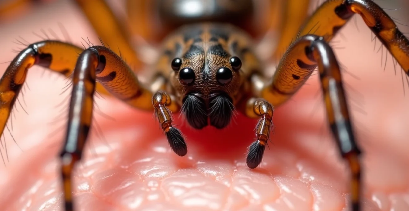
Physical characteristics of brown recluse spiders: Violin-Shaped dorsal markings
The most recognisable feature of adult brown recluse spiders is the distinctive violin-shaped marking on their cephalothorax, earning them alternative names such as “violin spider” or “fiddleback spider”. This dark brown to black marking appears with the violin’s neck pointing toward the spider’s abdomen. However, relying solely on this marking for identification can prove problematic, as juvenile specimens and recently moulted adults may display faded or absent markings. Additionally, the violin pattern’s intensity varies considerably among individuals and can be obscured by dirt or debris.
More reliable identification features include the spider’s unique eye arrangement, consisting of six eyes arranged in three pairs rather than the typical eight-eye configuration found in most spider species. These eyes form a semicircular pattern when viewed from above, providing a more consistent identification marker than dorsal markings. The legs appear uniformly coloured without distinct banding or markings, and the entire body lacks spines or dense hair coverage, giving brown recluses their characteristically smooth appearance.
Endemic regions: midwest and South-Central united states habitat zones
Brown recluse spiders demonstrate a remarkably specific geographic distribution, with established populations concentrated primarily in the south-central and midwestern United States. The core endemic range includes Arkansas, Kansas, Missouri, Oklahoma, Tennessee, and Texas, with smaller populations extending into adjacent areas of Alabama, Georgia, Illinois, Indiana, Iowa, Kentucky, Louisiana, Mississippi, Nebraska, and Ohio. This distribution pattern corresponds closely with specific climatic conditions and habitat requirements essential for population establishment and maintenance.
Understanding these geographic boundaries proves crucial for medical professionals, as confirmed brown recluse bites outside endemic areas should raise suspicion for alternative diagnoses or transportation-related exposure. The spiders’ inability to establish sustainable populations in regions with harsh winters or excessive humidity limits their natural spread, though isolated specimens occasionally appear in non-endemic areas through human transportation of infested materials.
Differential diagnosis from loxosceles deserta and other recluse species
Several Loxosceles species inhabit North America, requiring careful differentiation for accurate risk assessment and clinical management. The desert recluse ( Loxosceles deserta ) occupies southwestern regions including Arizona, California, Nevada, and Utah, typically appearing larger and lighter in colouration than its brown cousin. These species share similar venom compositions and clinical presentations, making geographic context essential for species identification.
Other recluse species, including the Arizona recluse ( Loxosceles arizonica ) and various South American species occasionally imported with cargo, present similar morphological characteristics but different distribution patterns. The mediterranean recluse ( Loxosceles rufescens ) has established limited populations in certain urban areas, particularly in California and Florida, though encounters remain relatively uncommon compared to native species.
Seasonal activity patterns and indoor nesting preferences
Brown recluse activity patterns follow predictable seasonal cycles that influence bite risk throughout the year. These spiders demonstrate peak activity during warmer months, typically from April through October, when hunting and reproductive behaviours intensify. During winter months, brown recluses enter a state of reduced activity called diapause, seeking shelter in protected locations where temperatures remain relatively stable.
Indoor environments provide ideal habitats for brown recluse establishment, offering consistent temperatures, abundant hiding places, and potential food sources. Common indoor nesting sites include undisturbed storage areas, closets, attics, basements, and spaces behind furniture or appliances. The spiders show particular preference for cardboard boxes, clothing piles, and areas with accumulated debris, where they can construct irregular webs for shelter rather than prey capture.
Cytotoxic venom composition and sphingomyelinase D mechanisms
The pathological effects of brown recluse envenomation result from a complex mixture of enzymatic proteins, with sphingomyelinase D serving as the primary toxic component responsible for tissue necrosis and systemic complications. This enzyme, unique to Loxosceles species, demonstrates remarkable potency despite the relatively small volume of venom injected during a bite. Understanding the biochemical mechanisms underlying tissue damage provides insight into the progressive nature of brown recluse envenomation and explains the delayed onset of severe symptoms.
The venom’s cytotoxic properties manifest through multiple pathways, including direct cellular damage, inflammatory cascade activation, and disruption of normal haemostatic mechanisms. Unlike neurotoxic venoms that primarily affect nerve transmission, brown recluse venom targets cellular membranes, blood vessel integrity, and immune system responses, creating a cascade of pathological changes that can progress over days to weeks following initial exposure.
Phospholipase D enzyme pathophysiology in dermal tissue
Sphingomyelinase D, also known as phospholipase D in some literature, represents the most thoroughly studied component of brown recluse venom. This enzyme cleaves sphingomyelin, a major component of cellular membranes, particularly abundant in red blood cell and endothelial cell membranes. The enzymatic action results in membrane instability, cellular lysis, and release of inflammatory mediators that propagate tissue damage beyond the immediate bite site.
The enzyme’s activity explains the characteristic progression of brown recluse wounds, beginning with localised cellular damage and gradually expanding as inflammatory responses amplify the initial injury. Dermal tissues show particular susceptibility to phospholipase D activity, with endothelial cells lining blood vessels representing primary targets for enzyme action. This selective vulnerability accounts for the vascular compromise and subsequent ischaemic necrosis observed in severe envenomations.
Complement cascade activation and inflammatory response pathways
Brown recluse venom triggers robust activation of the complement system, a crucial component of innate immunity that, when dysregulated, contributes significantly to tissue damage. Complement activation occurs through both classical and alternative pathways, generating potent inflammatory mediators including C3a and C5a anaphylatoxins. These complement fragments promote vasodilation, increased vascular permeability, and recruitment of inflammatory cells to the envenomation site.
The inflammatory response initiated by complement activation creates a self-perpetuating cycle of tissue damage, as recruited neutrophils release additional cytotoxic substances including reactive oxygen species and proteolytic enzymes. This inflammatory amplification explains why brown recluse wounds often worsen over the first several days following the bite, even in the absence of secondary bacterial infection.
Platelet aggregation and coagulation factor disruption
Venom-induced alterations in normal haemostatic mechanisms contribute to the complex pathophysiology of brown recluse envenomation. Sphingomyelinase D activity promotes platelet aggregation and adhesion to damaged vessel walls, potentially leading to thrombosis within affected tissues. Simultaneously, the enzyme disrupts normal coagulation factor function, creating a paradoxical situation where both thrombosis and bleeding tendencies may coexist.
These haemostatic disturbances manifest clinically as areas of tissue ischaemia due to microvascular thrombosis, while systemic effects may include prolonged bleeding times and increased susceptibility to haemorrhage. The balance between prothrombotic and anticoagulant effects varies among individuals and correlates with envenomation severity, explaining the wide spectrum of clinical presentations observed following brown recluse bites.
Hyaluronidase activity and tissue permeability enhancement
Hyaluronidase represents another significant component of brown recluse venom, functioning as a “spreading factor” that enhances the penetration and distribution of other toxic components. This enzyme degrades hyaluronic acid, a major component of the extracellular matrix that normally provides structural support and regulates tissue permeability. Hyaluronidase activity allows venom components to spread more readily through tissue planes, extending the zone of injury beyond the immediate bite location.
The enzyme’s action contributes to the characteristic gravitational spread observed in some brown recluse envenomations, where tissue damage progresses downward along anatomical planes due to gravity’s influence on venom distribution. This spreading pattern, combined with the progressive nature of tissue necrosis, explains why brown recluse wounds may continue expanding for days following the initial bite, particularly when located on dependent body parts.
Initial clinical presentation within 24-hour Post-Exposure window
The first 24 hours following brown recluse envenomation present unique diagnostic challenges due to the often subtle and non-specific nature of initial symptoms. Unlike many other arthropod bites that produce immediate pain and obvious inflammatory changes, brown recluse bites frequently begin with minimal discomfort, leading to delayed recognition and potential underestimation of injury severity. This characteristic presentation, termed the “painless bite phenomenon,” occurs because brown recluse fangs are extremely small and the initial venom injection may not immediately stimulate pain receptors.
Most patients report becoming aware of the bite only after noticing skin changes or experiencing delayed onset discomfort, typically 2-8 hours post-exposure. The initial bite site may appear as nothing more than a small red mark or slight swelling, easily dismissed as a minor skin irritation. However, careful observation during this critical period often reveals subtle signs that suggest significant envenomation, including the development of central pallor, surrounding erythema, and gradual onset of localised discomfort.
The absence of immediate severe symptoms frequently leads to delayed medical consultation, potentially missing the optimal window for early intervention strategies. Understanding this deceptive presentation proves crucial for both healthcare providers and individuals living in endemic areas, as early recognition can significantly influence clinical outcomes. Healthcare professionals should maintain a high index of suspicion for brown recluse envenomation when evaluating skin lesions in endemic regions, particularly when patients report finding spiders in their environment or discovering unexplained marks after sleeping or handling stored materials.
Temperature sensation at the bite site provides another early indicator of envenomation severity. Patients may report a burning or stinging sensation that develops gradually, often described as more intense than expected for such a small wound. This thermal dysesthesia, combined with progressive tenderness upon palpation, suggests active venom effects and warrants close monitoring for disease progression. The absence of immediate systemic symptoms during the first day should not provide false reassurance, as serious complications often develop 24-72 hours post-exposure.
Dermatological manifestations and progressive necrotic changes
The dermatological presentation of brown recluse envenomation evolves through predictable stages during the first 24 hours, with each phase providing important prognostic information about likely disease progression. Initial skin changes may appear subtle but demonstrate characteristic patterns that distinguish brown recluse bites from other arthropod injuries or infectious processes. Recognition of these early dermatological signs enables appropriate monitoring protocols and timely intervention when indicated.
Erythematous ring formation and central blanching patterns
Within 2-4 hours of envenomation, many brown recluse bites develop the classic “red, white, and blue” appearance that serves as an important diagnostic indicator. This tricolour pattern results from the complex vascular effects of sphingomyelinase D activity, creating concentric zones of different tissue responses. The central area often appears pale or blanched due to vasoconstriction and impaired circulation, while the surrounding tissue shows erythema from inflammatory vasodilation.
The outer zone may develop a bluish discolouration, particularly in more severe envenomations, indicating compromised tissue perfusion and early ischaemic changes. This colour progression provides valuable prognostic information, as bites that rapidly develop central blanching or cyanotic changes carry higher risk for subsequent necrosis. Photographic documentation of these colour changes proves valuable for monitoring progression and guiding treatment decisions.
Not all brown recluse bites develop the classic tricolour pattern, particularly in mild envenomations or when treatment begins early. Some bites may show only subtle erythema and swelling during the first day, making differentiation from other arthropod bites challenging. However, the presence of central blanching, even without the full tricolour pattern, should raise suspicion for brown recluse envenomation and prompt appropriate monitoring protocols.
Gravitational spread and dependent positioning effects
The anatomical location of brown recluse bites significantly influences the pattern and severity of tissue involvement, with dependent body parts showing increased risk for extensive damage. Bites on the lower extremities, particularly the thighs, calves, and ankles, often demonstrate gravitational spread of venom effects, creating elongated areas of tissue involvement that follow fascial planes and dependent drainage patterns.
This gravitational influence results from the combined effects of hyaluronidase activity and normal tissue fluid dynamics, allowing venom components to migrate downward through tissue spaces. Patients may notice that skin changes extend beyond the original bite location, creating irregular patterns of erythema and induration that can span several centimetres. This spreading pattern distinguishes brown recluse envenomation from localised bacterial infections, which typically show more uniform circular expansion.
Bites on the torso and upper extremities generally show less dramatic spreading but may still demonstrate eccentric patterns of involvement influenced by anatomical structures and patient positioning during sleep. The dependency effect explains why brown recluse bites discovered upon awakening often show more extensive involvement than might be expected from a simple spider bite, particularly when the affected area remained in a dependent position overnight.
Bullae formation and vesicular eruption timeline
Blister formation represents another characteristic feature of brown recluse envenomation, typically appearing 6-12 hours post-exposure in moderate to severe cases. These bullae result from extensive dermal-epidermal separation caused by inflammatory oedema and direct cytotoxic effects on basement membrane structures. The blisters may appear as single large lesions or multiple smaller vesicles, depending on the distribution of venom effects and individual tissue responses.
The fluid within brown recluse-induced bullae often appears clear initially but may become haemorrhagic as vascular damage progresses. Blister formation provides important prognostic information, as extensive bullae formation during the first 24 hours correlates with increased risk for subsequent tissue necrosis. Healthcare providers should avoid rupturing these blisters unnecessarily, as the overlying skin provides natural protection for underlying damaged tissues.
The timeline of blister development helps differentiate brown recluse envenomation from other vesicular eruptions. While conditions such as herpes simplex or contact dermatitis may produce similar-appearing blisters, the rapid onset and progressive nature of brown recluse-induced bullae, combined with appropriate clinical context, suggests arachnid envenomation requiring specialised management approaches.
Early signs of loxoscelism and tissue ischaemia
The term “loxoscelism” describes the constellation of findings associated with Loxosceles envenomation, ranging from localised skin changes to life-threatening systemic complications. Early recognition of loxoscelism during the first 24 hours proves crucial for appropriate risk stratification and treatment planning. Localised loxoscelism presents primarily with progressive skin changes, while systemic lox
oscelism may progress to systemic disease with haemolytic complications, making early recognition essential for appropriate medical management.
During the first 24 hours, clinicians should assess for subtle signs of tissue ischaemia that may herald more extensive damage. These early indicators include progressive coolness of the affected area, delayed capillary refill in tissues surrounding the bite, and development of a firm, indurated quality to the involved skin. The affected area may feel different from surrounding normal tissue, often described by patients as “tight” or “woody” in consistency.
Careful palpation reveals important information about tissue viability and venom distribution. Areas showing early ischaemic changes may demonstrate decreased skin mobility and increased resistance to gentle pressure. The border between involved and uninvolved tissue often shows a distinct demarcation that becomes more pronounced as ischaemia progresses. Healthcare providers should mark these borders with a skin marker to facilitate monitoring of disease progression during subsequent evaluations.
Temperature gradients across the affected area provide additional prognostic information during early assessment. Digital thermometry or infrared temperature measurement may reveal subtle temperature differences that precede visible tissue changes. Areas showing persistent coolness compared to surrounding skin carry higher risk for subsequent necrosis, particularly when combined with other indicators of compromised circulation such as delayed capillary refill or cyanotic discolouration.
Systemic symptomatology and haemolytic complications
While localised skin changes dominate the early presentation of brown recluse envenomation, systemic symptoms may develop within the first 24 hours, particularly in children, elderly patients, or individuals with compromised immune systems. These systemic manifestations result from haemolytic activity of circulating venom components and inflammatory mediator release, creating potentially life-threatening complications that require immediate medical intervention. Understanding the spectrum of systemic loxoscelism proves crucial for appropriate risk assessment and treatment planning.
The haemolytic effects of brown recluse venom stem primarily from sphingomyelinase D activity on red blood cell membranes, leading to intravascular haemolysis and subsequent complications. This enzymatic destruction of erythrocytes releases haemoglobin into the circulation, potentially overwhelming the body’s normal clearance mechanisms and leading to haemoglobinuria, anaemia, and renal complications. The severity of haemolytic effects correlates with the amount of venom injected and individual patient susceptibility factors.
Early systemic symptoms often begin subtly, with patients reporting general malaise, fatigue, or mild nausea that may be attributed to anxiety about the spider bite rather than recognised as signs of envenomation. These non-specific symptoms can progress rapidly to more concerning manifestations including fever, chills, headache, and abdominal pain. The development of dark-coloured urine represents a particularly ominous sign indicating significant haemolysis and potential renal involvement requiring urgent medical evaluation.
Coagulation abnormalities may manifest during the first day following severe envenomation, presenting as prolonged bleeding from minor wounds, spontaneous bruising, or petechial rashes on dependent body parts. These findings suggest consumption of clotting factors and platelet dysfunction secondary to venom effects. Laboratory evaluation during this period typically reveals thrombocytopaenia, prolonged coagulation times, and evidence of disseminated intravascular coagulation in the most severe cases.
Neurological symptoms, while less common during the acute phase, may include headache, dizziness, weakness, and altered mental status in severe systemic envenomations. These manifestations likely result from anaemia, metabolic disturbances, and inflammatory mediator effects rather than direct neurotoxic venom activity. Patients may also report muscle aches and joint pain, creating a syndrome resembling viral illness that can complicate diagnostic assessment.
Cardiovascular manifestations during the first 24 hours may include tachycardia, hypotension, and signs of volume depletion secondary to third-spacing of fluids and haemolytic complications. These findings indicate significant systemic involvement requiring intensive monitoring and supportive care. The combination of haemolysis, coagulopathy, and cardiovascular instability represents a medical emergency requiring immediate hospitalisation and specialised treatment protocols.
Emergency assessment protocols and first-day monitoring requirements
Effective management of suspected brown recluse envenomation during the critical first 24 hours requires systematic assessment protocols and vigilant monitoring for both local and systemic disease progression. Healthcare providers must balance the need for comprehensive evaluation with the recognition that many brown recluse bites resolve without significant complications. The challenge lies in identifying those cases requiring intensive monitoring while avoiding unnecessary alarm for patients with mild envenomation.
Initial assessment should begin with a detailed history focusing on the circumstances of the suspected bite, timing of symptom onset, and progression of local changes. Patients should be questioned about finding spiders in their environment, activities that might have led to spider contact, and any previous experiences with arthropod bites. Geographic considerations prove crucial, as confirmed brown recluse bites outside endemic areas require additional diagnostic consideration or evidence of transportation from endemic regions.
Physical examination during the first day should include careful inspection and documentation of the suspected bite site, including photographic records when possible. Measurement of affected areas, assessment of colour changes, and evaluation of tissue consistency provide baseline information for monitoring progression. The examination should extend beyond the immediate bite area to assess for regional lymphadenopathy, skin temperature variations, and signs of spreading inflammation or infection.
Vital sign monitoring becomes particularly important when systemic symptoms are present or suspected. Temperature elevation may indicate inflammatory responses or secondary bacterial infection, while tachycardia and hypotension suggest haemolytic complications or volume depletion. Blood pressure should be monitored closely in patients showing signs of systemic envenomation, as cardiovascular instability may develop rapidly in severe cases.
Laboratory evaluation during the first 24 hours should be tailored to the clinical presentation and severity of suspected envenomation. Patients with localised symptoms and no systemic manifestations may require only baseline studies, while those showing signs of systemic involvement need comprehensive haematological and biochemical assessment. Essential laboratory studies for suspected systemic loxoscelism include complete blood count with differential, comprehensive metabolic panel, coagulation studies, urinalysis, and lactate dehydrogenase levels as markers of haemolysis.
Monitoring protocols should establish clear parameters for reassessment and escalation of care. Patients with mild localised symptoms may be managed with outpatient follow-up at 24-48 hour intervals, while those showing signs of progression or systemic involvement require more intensive monitoring. Clear discharge instructions should include specific warning signs that warrant immediate medical reevaluation, such as expanding areas of skin involvement, development of systemic symptoms, or signs of secondary bacterial infection.
Communication with patients and families proves essential during the first day of care, as anxiety about potential complications can significantly impact the patient experience. Educational materials should explain the typical course of brown recluse envenomation, expected timeline for symptom development, and specific monitoring requirements. Patients should understand that while serious complications can occur, most brown recluse bites resolve without permanent damage when appropriately managed.
Documentation requirements during emergency assessment should include detailed descriptions of wound characteristics, photographic evidence when possible, and clear treatment plans with follow-up arrangements. This documentation proves valuable for continuity of care and medical-legal considerations, particularly given the diagnostic challenges associated with confirming brown recluse envenomation. Healthcare providers should maintain detailed records of patient education provided and specific instructions given for home monitoring and when to seek additional medical care.