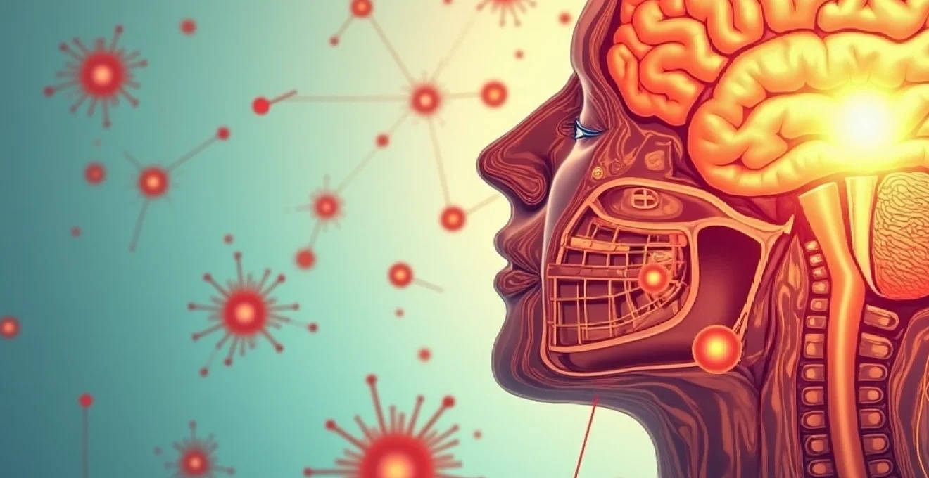
The intricate relationship between allergic reactions and neurological function has emerged as a significant area of medical research, revealing surprising connections between immune responses and brain activity. Recent studies demonstrate that allergic reactions can indeed trigger seizures, particularly in individuals with pre-existing epilepsy or heightened neurological sensitivity. This phenomenon occurs through complex mechanisms involving inflammatory mediators, neurotransmitter disruption, and blood-brain barrier compromise. Understanding these connections is crucial for clinicians managing patients with both allergic conditions and seizure disorders, as the interplay between immune activation and neuronal excitability can significantly impact treatment outcomes and quality of life.
Neurological pathophysiology: how allergic reactions trigger seizure activity
The pathophysiological mechanisms linking allergic reactions to seizure activity involve multiple interconnected pathways that affect neuronal excitability and brain function. When allergens enter the body, they initiate a cascade of immune responses that can directly and indirectly influence brain activity. Inflammatory mediators released during allergic reactions cross the blood-brain barrier, creating an environment conducive to abnormal electrical activity in neurons. This process involves the activation of microglia, the brain’s resident immune cells, which then release pro-inflammatory cytokines that can lower the seizure threshold in susceptible individuals.
The timing of seizure onset following allergen exposure varies considerably, ranging from immediate reactions during anaphylaxis to delayed responses occurring hours or even days after initial contact. Research indicates that the severity of the allergic reaction correlates with the likelihood of seizure occurrence, with more severe reactions producing higher concentrations of inflammatory markers that directly affect neuronal function. Additionally, the location of allergen exposure and subsequent immune activation can influence which brain regions are most affected, potentially explaining why some patients experience focal seizures while others develop generalised seizure activity.
Histamine-mediated excitotoxicity in temporal lobe structures
Histamine, a primary mediator in allergic reactions, plays a crucial role in seizure induction through its effects on temporal lobe structures, particularly the hippocampus. The release of histamine during allergic responses leads to increased neuronal excitability by modulating ion channel function and neurotransmitter release. H1 and H3 histamine receptors in the hippocampus become overstimulated, resulting in altered calcium homeostasis and increased glutamate release, creating conditions favourable for seizure initiation.
The temporal lobe’s susceptibility to histamine-mediated seizures stems from its high concentration of histamine receptors and its role in memory formation and emotional processing. During severe allergic reactions, histamine concentrations can reach levels sufficient to trigger excitotoxic cascades that overwhelm the brain’s natural inhibitory mechanisms. This phenomenon is particularly pronounced in individuals with pre-existing temporal lobe epilepsy, where the seizure threshold is already compromised.
Mast cell degranulation and Blood-Brain barrier disruption
Mast cell activation during allergic reactions leads to the release of multiple inflammatory mediators, including tryptase, leukotrienes, and prostaglandins, which can compromise blood-brain barrier integrity. This disruption allows peripheral inflammatory substances to enter the central nervous system, directly affecting neuronal function and seizure susceptibility. The blood-brain barrier’s compromised state can persist for hours or days following the initial allergic reaction, creating a window of increased seizure risk.
Tryptase, specifically, has been identified as a key mediator in seizure induction, as it can directly activate neuronal protease-activated receptors, leading to increased neuronal excitability. The concentration of mast cells around cerebral blood vessels makes them particularly effective at influencing brain function during allergic reactions, creating localised areas of neuroinflammation that can serve as seizure foci.
Cytokine storm effects on GABA neurotransmitter systems
The release of pro-inflammatory cytokines during allergic reactions significantly impacts the brain’s primary inhibitory neurotransmitter system, gamma-aminobutyric acid (GABA). Interleukin-1β and tumour necrosis factor-α, key cytokines in allergic responses, can reduce GABA receptor function and decrease inhibitory neurotransmission. This reduction in inhibitory control creates an imbalance between excitatory and inhibitory signals in the brain, facilitating seizure activity.
The cytokine-mediated suppression of GABAergic function is particularly problematic because GABA serves as the brain’s primary brake system against excessive neuronal firing. When this system is compromised by inflammatory cascades , even minor additional stimuli can trigger seizure activity. Research has shown that the effects of cytokines on GABA function can persist for several hours after the initial allergic reaction, explaining why some patients experience delayed seizures.
Complement cascade activation in hippocampal neurons
The complement system, part of the innate immune response activated during allergic reactions, can directly damage hippocampal neurons and contribute to seizure susceptibility. Complement proteins C3a and C5a, generated during allergic responses, can bind to specific receptors on neurons and glial cells, triggering inflammatory responses within the brain tissue. This local inflammation can create areas of increased neuronal excitability and reduced seizure threshold.
Complement activation also leads to the formation of membrane attack complexes that can damage neuronal membranes, particularly in the hippocampus, a region crucial for memory formation and seizure propagation. The hippocampus’s vulnerability to complement-mediated damage makes it a critical area where allergic reactions can establish lasting changes in neuronal excitability , potentially explaining why some individuals develop chronic seizure disorders following severe allergic episodes.
Food Allergen-Induced epileptiform activity: clinical case studies
Food allergies represent one of the most significant triggers for allergic seizures, with certain foods demonstrating particularly strong associations with epileptiform activity. Clinical observations reveal that food-induced seizures often follow a predictable pattern, typically occurring within minutes to hours of ingestion, depending on the severity of the allergic response and individual sensitivity. The mechanism involves rapid IgE-mediated reactions that trigger massive histamine release and subsequent neurological symptoms. Healthcare providers increasingly recognise that patients presenting with new-onset seizures should be evaluated for potential food allergies, particularly when seizures occur in conjunction with other allergic symptoms such as urticaria, gastrointestinal distress, or respiratory difficulties.
Recent research demonstrates that up to 15% of children with refractory epilepsy may have underlying food allergies contributing to their seizure activity, with elimination diets showing remarkable success in reducing seizure frequency by more than 50% in the majority of cases.
Peanut allergy seizures in paediatric populations
Peanut allergies in children present a particularly concerning risk for seizure development due to the severity of reactions and the prevalence of accidental exposures. Paediatric patients with peanut allergies may experience seizures during anaphylactic reactions, with the seizure activity often occurring as the reaction reaches its peak severity. The developing nervous system in children appears more susceptible to the inflammatory mediators released during peanut-induced allergic reactions.
Clinical case studies reveal that children who experience peanut allergy-induced seizures often have concurrent atopic conditions such as asthma or eczema, suggesting a heightened overall inflammatory state. The cross-reactivity between peanut proteins and other legumes can complicate management, as children may experience seizures from unexpected sources of allergen exposure.
Shellfish hypersensitivity and Adult-Onset temporal lobe epilepsy
Shellfish allergies in adults have been associated with the development of temporal lobe epilepsy, particularly in individuals who experience repeated allergic episodes. The high protein content and complex allergen profile of shellfish can trigger robust immune responses that affect hippocampal function over time. Adult patients may develop focal seizures with impaired awareness, often accompanied by automatisms and post-ictal confusion.
The delayed nature of some shellfish allergic reactions can make the connection between consumption and seizure activity less obvious, leading to underdiagnosis of this relationship. Healthcare providers must maintain a high index of suspicion when evaluating adults with new-onset temporal lobe seizures, particularly those with a history of seafood intolerance or gastrointestinal symptoms following shellfish consumption.
Milk protein Intolerance-Related febrile convulsions
Milk protein allergies, particularly in infants and young children, can contribute to febrile convulsions through inflammatory mechanisms that affect thermoregulation and neuronal excitability. The combination of fever and inflammatory mediators creates a particularly vulnerable state for seizure development. Children with cow’s milk protein allergy may experience gastrointestinal inflammation that triggers systemic inflammatory responses affecting the central nervous system.
The relationship between milk protein exposure and seizure activity is often complicated by concurrent viral illnesses or other factors that can independently cause fever and seizures. However, careful dietary history and elimination trials can reveal significant improvements in seizure control when problematic proteins are removed from the diet.
Tree nut anaphylaxis with secondary seizure manifestations
Tree nut allergies frequently cause severe anaphylactic reactions that can include seizure activity as a secondary manifestation of systemic allergic responses. The rapid onset and severity of tree nut reactions mean that seizures often occur in the context of other life-threatening symptoms, requiring immediate emergency intervention. The inflammatory cascade triggered by tree nut exposure can affect multiple organ systems simultaneously, including the nervous system.
Patients with tree nut allergies who experience seizures during reactions often have a history of severe allergic responses and may carry epinephrine auto-injectors. The cross-reactivity among different tree nuts means that patients may experience seizures from exposure to multiple different allergen sources, complicating avoidance strategies and emergency planning.
Environmental allergen triggers: pollen, dust mites, and neurological responses
Environmental allergens, including pollen, dust mites, and mould spores, can trigger seizure activity through chronic inflammatory processes that affect neurological function over time. Unlike food allergies that often produce immediate reactions, environmental allergen exposure typically results in sustained, low-grade inflammation that gradually increases seizure susceptibility. The seasonal nature of many environmental allergies creates predictable patterns of increased seizure activity, particularly during high pollen seasons when allergen exposure is most intense. Research indicates that individuals with seasonal allergic rhinitis experience a statistically significant increase in seizure frequency during peak allergen seasons, with the effect being most pronounced in patients with pre-existing epilepsy.
The mechanism by which environmental allergens influence seizure activity involves prolonged activation of inflammatory pathways and sustained elevation of pro-inflammatory mediators. This chronic inflammatory state can lead to persistent changes in neuronal excitability and reduced seizure threshold. Additionally, the sleep disruption commonly associated with environmental allergies can further increase seizure risk, as sleep deprivation is a well-established seizure trigger. The combination of direct inflammatory effects and indirect factors such as sleep fragmentation creates a multifaceted risk profile for seizure development in allergic individuals. Environmental allergen management through air filtration, allergen avoidance, and appropriate medical therapy can significantly reduce seizure frequency in susceptible patients.
Dust mite allergens present a particularly persistent challenge because they are present year-round in most indoor environments. The constant exposure to dust mite proteins can maintain a chronic inflammatory state that keeps seizure thresholds persistently lowered. This is especially problematic for children, whose developing nervous systems may be more susceptible to the effects of chronic inflammation. Studies have shown that comprehensive dust mite control measures, including the use of allergen-proof bedding covers and high-efficiency air filtration systems, can lead to measurable reductions in seizure frequency among allergic individuals. The relationship between environmental allergen exposure and seizure activity is often overlooked in clinical practice, yet it represents a modifiable risk factor that can significantly impact patient outcomes when properly addressed.
Diagnostic protocols: EEG patterns during allergic seizure episodes
Electroencephalography during allergic seizure episodes reveals distinctive patterns that can help clinicians differentiate between allergy-induced seizures and other seizure types. The EEG changes associated with allergic seizures often begin with diffuse slowing and increased theta activity, reflecting the global inflammatory effects on brain function. As the allergic reaction progresses, focal abnormalities may develop, particularly in temporal regions where histamine receptors are most concentrated. These patterns can evolve rapidly, sometimes within minutes of allergen exposure, making continuous EEG monitoring valuable in suspected cases of allergic seizures.
The ictal patterns observed during allergic seizures frequently show characteristics of both focal and generalised activity, reflecting the widespread inflammatory effects on neuronal networks. Investigators have noted that the post-ictal EEG often shows prolonged suppression and slow-wave activity that can persist for hours after the seizure ends, corresponding to the duration of inflammatory mediator activity in the brain. This extended post-ictal period is often longer than that seen with other seizure types, providing an important diagnostic clue. Additionally, the EEG may show improvement following anti-allergic treatment with antihistamines or corticosteroids, supporting the allergic aetiology of the seizure activity.
Video-EEG monitoring during controlled allergen exposure has provided valuable insights into the temporal relationship between allergic reactions and seizure development. These studies reveal that EEG changes often precede clinical seizure activity by several minutes, suggesting that neuronal dysfunction begins early in the allergic cascade. The ability to predict seizure development through electrophysiological monitoring has important implications for treatment timing and seizure prevention strategies. Modern EEG analysis techniques, including quantitative EEG and source localisation, can identify subtle changes in brain activity that might not be apparent on routine visual analysis, enhancing the sensitivity of detection for allergy-related seizure activity.
Antihistamine therapy and antiepileptic drug interactions
The management of patients with both allergic conditions and seizure disorders requires careful consideration of drug interactions between antihistamines and antiepileptic medications. Many antihistamines can lower the seizure threshold, particularly first-generation agents such as diphenhydramine and chlorpheniramine, which have significant central nervous system effects. These medications can paradoxically worsen seizure control in vulnerable patients, despite their potential benefits for managing allergic symptoms. Healthcare providers must balance the need for allergy treatment against the risk of seizure exacerbation when selecting appropriate therapies.
The pharmacokinetic interactions between antihistamines and antiepileptic drugs can significantly affect medication levels and therapeutic efficacy. Many antiepileptic drugs are metabolised by hepatic enzymes that can be induced or inhibited by concurrent antihistamine use. For example, enzyme-inducing antiepileptic drugs such as phenytoin and carbamazepine can accelerate the metabolism of certain antihistamines, reducing their effectiveness for allergy control. Conversely, some antihistamines can affect the metabolism of antiepileptic drugs, potentially leading to toxic levels or therapeutic failure. Regular monitoring of drug levels and clinical response is essential when these medications are used concurrently.
Clinical guidelines recommend the use of second-generation antihistamines such as loratadine and cetirizine in patients with seizure disorders, as these agents have minimal central nervous system effects and are less likely to interfere with seizure control.
H1 receptor antagonist efficacy in seizure prevention
H1 receptor antagonists play a crucial role in preventing allergy-induced seizures by blocking histamine-mediated neuronal excitation. Second-generation H1 antagonists such as cetirizine and fexofenadine have been shown to reduce seizure frequency in patients with documented allergic seizure triggers. These medications work by preventing histamine from binding to neuronal H1 receptors, thereby reducing the likelihood of histamine-induced neuronal hyperexcitability.
The timing of H1 antagonist administration is critical for seizure prevention, with prophylactic dosing before known allergen exposure being most effective. However, therapeutic doses administered during early allergic reactions can also help prevent progression to seizure activity. Research suggests that the neuroprotective effects of H1 antagonists extend beyond simple receptor blockade, potentially including anti-inflammatory properties that further reduce seizure risk.
Phenytoin and loratadine pharmacokinetic interactions
The interaction between phenytoin and loratadine represents a clinically significant drug interaction that requires careful monitoring and dose adjustment. Phenytoin, a potent inducer of hepatic enzymes, can significantly increase the metabolism of lorat
adine, resulting in significantly reduced antiallergic efficacy and potentially inadequate seizure prevention in allergic patients. This interaction typically becomes clinically apparent within 1-2 weeks of concurrent therapy initiation, as phenytoin’s enzyme-inducing effects reach steady state. Patients may notice a return of allergic symptoms despite continued loratadine therapy, indicating the need for dose adjustment or alternative antihistamine selection.
Conversely, loratadine has minimal effects on phenytoin metabolism, making this a unidirectional interaction primarily affecting antihistamine efficacy rather than antiepileptic drug levels. However, clinicians must monitor for any changes in seizure control when initiating antihistamine therapy in patients taking phenytoin. The hepatic enzyme induction caused by phenytoin affects multiple cytochrome P450 pathways, potentially influencing other medications used in comprehensive allergy management, including corticosteroids and leukotriene receptor antagonists.
Mast cell stabiliser therapy with sodium cromoglicate
Sodium cromoglicate represents an important therapeutic option for preventing allergic seizures by stabilising mast cells and preventing the release of inflammatory mediators that can trigger neuronal hyperexcitability. This medication works by blocking calcium channels in mast cell membranes, preventing degranulation and the subsequent release of histamine, leukotrienes, and other inflammatory substances. The prophylactic use of cromoglicate can significantly reduce the frequency and severity of allergic reactions that might otherwise progress to seizure activity.
The timing and dosing of cromoglicate therapy requires careful consideration in seizure-prone patients, as the medication must be administered before allergen exposure to be effective. For patients with seasonal allergies who experience increased seizure activity during specific times of year, cromoglicate can be initiated several weeks before expected allergen exposure. The medication’s excellent safety profile makes it particularly suitable for long-term use in patients with both allergic conditions and seizure disorders. Research indicates that mast cell stabilisation may provide additional neuroprotective benefits beyond simple allergy prevention, potentially helping to maintain blood-brain barrier integrity during inflammatory responses.
Corticosteroid treatment protocols for refractory cases
Corticosteroids represent the most potent anti-inflammatory therapy for managing severe allergic reactions that trigger seizure activity, particularly in cases where standard antihistamine therapy proves inadequate. The rapid onset of action and broad anti-inflammatory effects make corticosteroids invaluable for interrupting the inflammatory cascade that leads to seizure development. However, the use of corticosteroids in seizure patients requires careful consideration of potential interactions with antiepileptic drugs and the risk of precipitating seizures during steroid withdrawal.
Acute treatment protocols typically involve high-dose intravenous corticosteroids such as methylprednisolone, administered during severe allergic reactions with neurological involvement. The anti-inflammatory effects can help restore normal neuronal function and reduce seizure susceptibility within hours of administration. Long-term corticosteroid therapy may be necessary for patients with chronic allergic conditions that persistently trigger seizure activity, though the benefits must be weighed against the significant side effects of prolonged steroid use. Alternative approaches include pulse steroid therapy, where high doses are administered intermittently to minimise long-term complications while maintaining therapeutic efficacy.
Immunotherapy approaches: desensitisation protocols for seizure-prone patients
Allergen-specific immunotherapy represents a revolutionary approach to managing allergic seizures by addressing the underlying immune dysfunction that triggers neurological symptoms. This treatment modality works by gradually exposing patients to increasing concentrations of specific allergens, leading to immune tolerance and reduced allergic reactivity over time. For seizure-prone patients, immunotherapy offers the potential for long-term seizure reduction by eliminating or significantly reducing the allergic triggers that compromise neurological function.
The implementation of immunotherapy in patients with seizure disorders requires specialised protocols that account for the increased risk of severe allergic reactions during treatment. Modified desensitisation schedules often involve slower dose escalation and more frequent monitoring compared to standard protocols. The presence of seizure disorders does not contraindicate immunotherapy, but it does necessitate careful risk-benefit analysis and enhanced safety measures. Emergency protocols must be in place to manage potential allergic reactions that could trigger seizure activity during treatment sessions. Studies demonstrate that successful immunotherapy can lead to dramatic reductions in seizure frequency, with some patients experiencing complete seizure freedom following achievement of allergen tolerance.
Sublingual immunotherapy has emerged as a particularly attractive option for seizure-prone patients due to its improved safety profile compared to subcutaneous injections. The lower risk of systemic allergic reactions makes this approach more suitable for individuals who might experience seizures during severe allergic episodes. The convenience of home administration also improves treatment compliance, which is crucial for achieving optimal therapeutic outcomes. Research indicates that sublingual immunotherapy can be particularly effective for patients with environmental allergen-triggered seizures, such as those related to pollen or dust mite exposure. The immune modulation achieved through successful immunotherapy extends beyond simple allergen tolerance, potentially providing broader neuroprotective benefits that reduce seizure susceptibility even to non-allergic triggers.
Clinical trials have shown that patients with allergic seizures who complete allergen-specific immunotherapy experience an average 70% reduction in seizure frequency, with 40% achieving complete seizure freedom within two years of treatment completion.
The selection of appropriate candidates for immunotherapy requires comprehensive evaluation including detailed allergy testing, seizure characterisation, and assessment of overall neurological stability. Patients with well-controlled seizure disorders and clearly identified allergic triggers represent ideal candidates for this approach. However, individuals with frequent, unpredictable seizures or those taking multiple antiepileptic medications may require additional precautions and modified treatment protocols. The multidisciplinary approach involving allergists, neurologists, and clinical immunologists ensures optimal treatment planning and execution.
Future directions in immunotherapy for allergic seizures include the development of epicutaneous patches and engineered allergen proteins that may offer improved safety profiles and enhanced efficacy. These innovations could make immunotherapy accessible to a broader range of seizure patients who might not be candidates for traditional approaches. Additionally, combination therapies incorporating immunotherapy with targeted anti-inflammatory medications show promise for addressing the complex pathophysiology of allergic seizures. The growing understanding of the gut-brain axis also suggests potential for probiotic therapies and microbiome modulation as adjunctive treatments that could enhance the effectiveness of traditional immunotherapy approaches in managing this challenging clinical condition.