Ureaplasma infections represent a significant yet often overlooked factor in reproductive health challenges affecting millions of couples worldwide. These microscopic bacteria, particularly Ureaplasma urealyticum and Ureaplasma parvum , inhabit the urogenital tract of 40-80% of sexually active individuals, frequently remaining silent until fertility problems emerge. Unlike conventional bacterial infections that announce their presence through obvious symptoms, Ureaplasma operates as a stealth pathogen, potentially compromising reproductive function through subtle inflammatory processes that can persist for years undetected.
The relationship between Ureaplasma colonisation and infertility challenges traditional medical paradigms. While these organisms are commonly dismissed as harmless commensals, emerging research reveals their capacity to trigger chronic inflammatory responses that significantly impact both male and female fertility. Recent studies indicate that Ureaplasma infections may contribute to up to 30% of unexplained infertility cases, highlighting the urgent need for comprehensive screening and targeted treatment protocols in reproductive medicine.
Ureaplasma urealyticum and ureaplasma parvum: pathogenic mechanisms in reproductive health
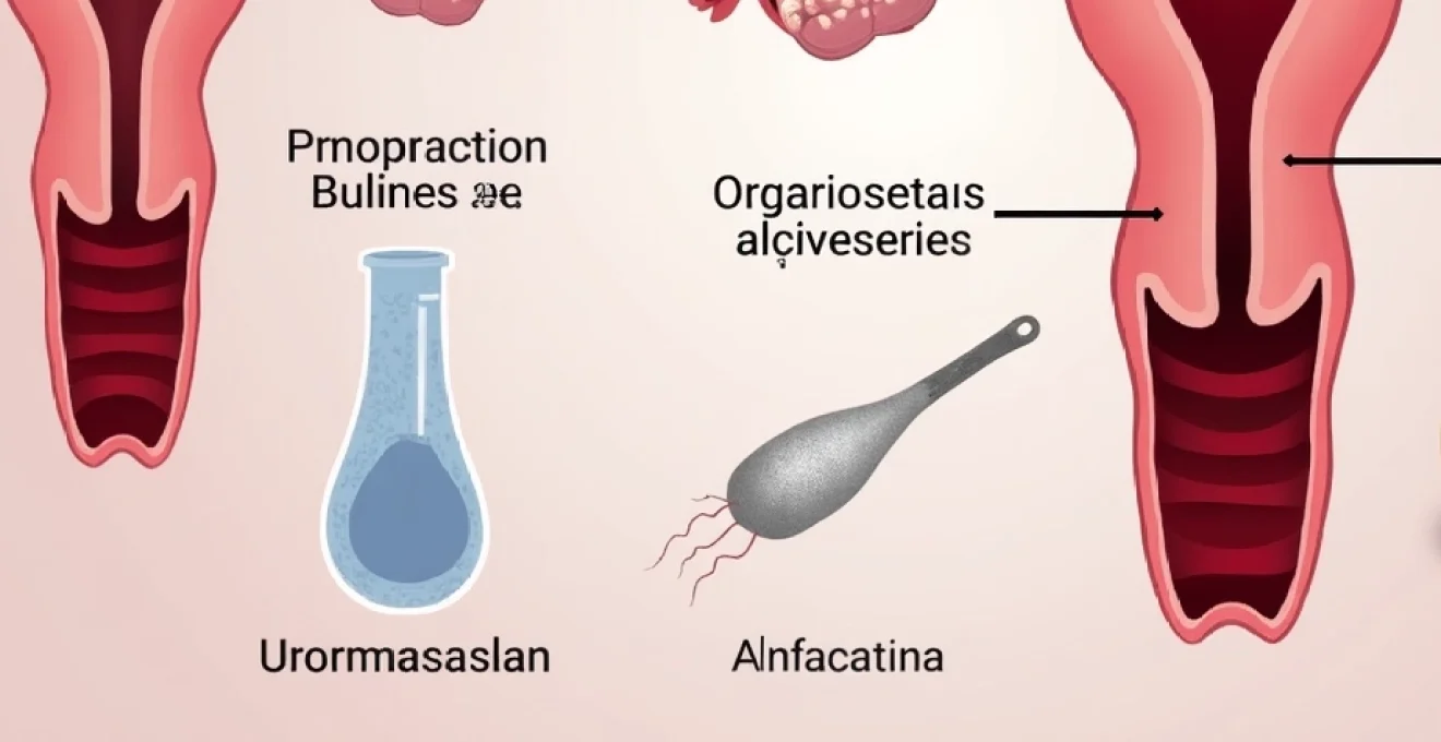
Understanding how Ureaplasma species compromise fertility requires examining their unique biological characteristics and pathogenic strategies. These bacteria belong to the Mollicutes class, distinguished by their absence of a cell wall and remarkably small genome size. This structural peculiarity allows them to evade conventional antibiotic treatments while maintaining their ability to colonise and invade reproductive tissues with remarkable efficiency.
Bacterial adhesion to urogenital epithelial cells and tissue invasion
Ureaplasma species demonstrate sophisticated adhesion mechanisms that enable them to establish persistent colonies within the urogenital tract. The bacteria utilise specialised surface proteins, including multiple-banded antigens (MBA), to attach to urethral epithelial cells, spermatozoa, and even erythrocytes. Research indicates that these adhesion receptors specifically target sialyl residues and sulfated compounds on host cell surfaces, creating stable bacterial-host interactions that resist natural clearance mechanisms.
Once established, Ureaplasma can migrate from the lower genital tract to sterile environments such as the endometrium and fallopian tubes. This ascending infection pattern occurs in approximately 40% of colonised women, with Ureaplasma parvum showing particular affinity for upper reproductive tract colonisation. The bacteria’s ability to penetrate tissue barriers and establish chronic infections in typically sterile anatomical sites represents a critical factor in their fertility-compromising potential.
Inflammatory cytokine release and immune system dysregulation
Ureaplasma infections trigger complex inflammatory cascades that fundamentally alter the reproductive tract’s immunological environment. The bacteria stimulate the production of pro-inflammatory cytokines, including interleukin-1β, interleukin-6, and tumour necrosis factor-α, creating a hostile microenvironment for gamete function and embryo development. This inflammatory response can persist for months or years, even in the absence of obvious symptoms.
The chronic nature of Ureaplasma-induced inflammation particularly affects endometrial receptivity and fallopian tube function. Studies demonstrate that infected tissues exhibit elevated levels of inflammatory markers that interfere with normal reproductive processes. The sustained immune activation creates a paradoxical situation where the body’s defence mechanisms actually contribute to fertility impairment , highlighting the complex interplay between infection, immunity, and reproductive success.
Sperm DNA fragmentation and motility impairment pathways
Male fertility suffers significantly under Ureaplasma colonisation through direct effects on sperm quality and function. The bacteria attach to sperm membranes, creating physical impediments to normal motility while simultaneously inducing oxidative stress that damages sperm DNA. Research reveals that Ureaplasma-positive semen samples show significantly higher levels of reactive oxygen species (ROS) and malondialdehyde, both indicators of oxidative cellular damage.
The molecular mechanisms underlying sperm impairment involve multiple pathways. Ureaplasma reduces expression of P34H protein, essential for sperm-zona pellucida interaction, while simultaneously decreasing hyaluronidase activity required for the acrosome reaction. These biochemical alterations result in measurable decreases in sperm concentration, progressive motility, and normal morphology percentages, directly correlating with reduced fertilisation potential.
Endometrial receptivity alterations through chronic inflammation
Female reproductive success depends critically on optimal endometrial receptivity during the implantation window. Ureaplasma infections disrupt this delicate process through persistent inflammatory changes that alter endometrial architecture and molecular signalling. Chronic endometritis, often associated with Ureaplasma colonisation, creates an environment hostile to embryo implantation and early pregnancy development.
The inflammatory mediators released during Ureaplasma infection interfere with normal hormonal signalling pathways essential for endometrial preparation. This disruption manifests as altered gene expression patterns, compromised vascular development, and abnormal immune cell populations within the endometrium. Consequently, even successfully fertilised embryos may fail to implant or may experience early pregnancy loss due to the hostile uterine environment created by chronic bacterial colonisation.
Clinical evidence linking ureaplasma infections to male factor infertility
The body of clinical evidence supporting Ureaplasma’s role in male infertility continues to expand, with numerous studies demonstrating significant correlations between bacterial presence and compromised sperm parameters. Meta-analyses examining thousands of patients reveal consistent patterns of reduced fertility potential among Ureaplasma-positive men, providing compelling evidence for the bacteria’s pathogenic role in reproductive dysfunction.
Seminal fluid analysis abnormalities in Ureaplasma-Positive patients
Comprehensive seminal fluid analyses consistently reveal distinctive abnormalities in men with Ureaplasma colonisation. Infected patients demonstrate significantly reduced sperm concentrations, with average decreases of 25-40% compared to uninfected controls. Progressive motility, perhaps the most critical parameter for natural conception, shows even more dramatic impairment, with some studies reporting reductions exceeding 50% in Ureaplasma-positive samples.
Morphological assessments reveal that Ureaplasma presence correlates with increased percentages of abnormally shaped sperm. The bacteria’s attachment to sperm surfaces appears to interfere with normal cellular development processes, resulting in head defects, midpiece abnormalities, and tail malformations. These morphological changes, combined with reduced motility, create a compound effect that significantly diminishes the probability of successful fertilisation through natural conception or assisted reproductive technologies.
Laboratory investigations consistently demonstrate that polymorphonuclear elastase levels increase substantially in Ureaplasma-positive semen samples, indicating ongoing inflammatory processes that further compromise sperm function and survival.
Epidemiological studies on ureaplasma prevalence in infertile men
Large-scale epidemiological investigations reveal striking differences in Ureaplasma prevalence between fertile and infertile male populations. Studies across diverse geographical regions consistently report detection rates of 5-58% in infertile men compared to 3-31% in proven fertile controls. These prevalence differentials suggest a clear association between bacterial colonisation and impaired reproductive capacity.
Particularly compelling evidence emerges from studies examining Ureaplasma urealyticum specifically, which shows stronger correlations with infertility than its counterpart, Ureaplasma parvum . Geographic variations in prevalence rates reflect differences in sexual behaviour patterns, screening practices, and antibiotic usage, but the consistent trend toward higher detection rates in infertile populations remains remarkably stable across different study populations and methodologies.
Correlation between ureaplasma load and sperm parameter deterioration
Quantitative PCR studies demonstrate clear dose-response relationships between Ureaplasma bacterial loads and the severity of sperm parameter abnormalities. Men with high bacterial concentrations (>10^4 colony-forming units per millilitre) exhibit more severe impairments across multiple sperm quality metrics compared to those with lower bacterial loads. This correlation suggests that bacterial burden, rather than mere presence, determines the extent of reproductive dysfunction.
The relationship between bacterial load and fertility impairment extends beyond simple sperm counts to encompass functional parameters such as DNA integrity and fertilisation capacity. High Ureaplasma loads correlate with increased DNA fragmentation indices, often exceeding 30% in severely affected patients compared to normal values below 15%. This DNA damage represents a critical factor in both natural conception difficulties and reduced success rates in assisted reproductive procedures.
Impact on acrosome reaction and Sperm-Oocyte binding capacity
Advanced reproductive technologies have enabled detailed examination of Ureaplasma’s effects on specific sperm functions essential for fertilisation. The bacteria significantly impair the acrosome reaction, the process by which sperm release enzymes necessary for penetrating the egg’s protective layers. Ureaplasma-infected sperm show reduced hyaluronidase activity and compromised calcium channel function, both critical for successful acrosome reactions.
Sperm-oocyte binding studies reveal that Ureaplasma presence substantially reduces the number of sperm capable of binding to the zona pellucida. This reduction stems from altered surface protein expression and compromised membrane integrity resulting from bacterial attachment and inflammatory responses. The cumulative effect of these molecular-level disruptions translates into measurably reduced fertilisation rates in both natural conception attempts and in vitro fertilisation procedures.
Ureaplasma-associated female reproductive complications and fertility outcomes
Female reproductive health faces multifaceted challenges from Ureaplasma colonisation, with infections affecting every level of the reproductive tract from the vagina to the fallopian tubes. The bacteria’s ability to ascend from lower genital tract colonisation sites to sterile upper reproductive tract environments creates opportunities for chronic inflammatory conditions that significantly compromise fertility potential.
Pelvic inflammatory disease development and fallopian tube scarring
Ureaplasma represents a significant risk factor for pelvic inflammatory disease (PID) development, particularly when bacterial loads exceed normal commensal levels. Unlike acute PID caused by conventional pathogens, Ureaplasma-associated inflammation often follows a subclinical course, with patients experiencing minimal symptoms while substantial tissue damage accumulates over time. This “silent” progression makes early detection and intervention challenging, often resulting in irreversible reproductive tract damage.
Fallopian tube scarring represents one of the most serious consequences of chronic Ureaplasma infection. The bacteria’s inflammatory mediators trigger fibroblast proliferation and collagen deposition within tube walls, leading to luminal narrowing, adhesion formation, and complete tubal occlusion in severe cases. Studies indicate that up to 15% of women with chronic Ureaplasma infections develop some degree of tubal factor infertility, with bilateral tubal blockage occurring in approximately 5% of chronically infected patients.
The progressive nature of Ureaplasma-induced tubal damage means that early colonisation events can have long-term consequences for fertility potential. Even after successful bacterial eradication, existing scar tissue and adhesions typically persist, requiring surgical intervention or assisted reproductive technologies to overcome mechanical barriers to conception . This reality underscores the importance of early detection and treatment before irreversible structural damage occurs.
Endometritis-related implantation failure mechanisms
Chronic endometritis associated with Ureaplasma colonisation creates a hostile uterine environment that significantly impairs embryo implantation success rates. The condition affects 25-30% of women with chronic Ureaplasma infections, often remaining asymptomatic until fertility challenges prompt comprehensive evaluation. Endometrial biopsies from affected patients reveal characteristic inflammatory infiltrates, disrupted glandular architecture, and altered molecular expression patterns that interfere with normal implantation processes.
The molecular mechanisms underlying implantation failure involve disrupted expression of critical adhesion molecules, altered prostaglandin synthesis, and abnormal immune cell populations within the endometrium. Ureaplasma-induced inflammation particularly affects the expression of integrins and selectins essential for embryo-endometrial interactions. Additionally, the chronic inflammatory state alters local hormone sensitivity, reducing the endometrium’s ability to respond appropriately to progesterone and other reproductive hormones.
Research demonstrates that women with chronic Ureaplasma endometritis experience implantation rates 40-50% lower than uninfected controls, highlighting the significant impact of bacterial colonisation on reproductive success.
Cervical factor infertility and mucus viscosity changes
Ureaplasma colonisation significantly alters cervical mucus production and composition, creating additional barriers to natural conception. The bacteria’s inflammatory effects disrupt normal hormonal regulation of cervical glands, resulting in mucus with altered pH, increased viscosity, and reduced sperm-penetrating capacity. These changes can persist even after bacterial loads decrease, suggesting that inflammatory damage to cervical tissue may have long-lasting effects on reproductive function.
Cervical mucus quality assessment reveals that Ureaplasma-positive women produce mucus with significantly reduced elasticity and spinnbarkeit compared to uninfected controls. The altered mucus composition creates a hostile environment for sperm survival and transport, with studies showing 60-70% reductions in sperm recovery from post-coital cervical mucus samples in infected women. These findings highlight how bacterial colonisation can impact fertility through multiple mechanisms simultaneously.
Recurrent pregnancy loss associations with chronic ureaplasma colonisation
Women experiencing recurrent pregnancy loss show substantially higher rates of Ureaplasma colonisation compared to those with successful pregnancy outcomes. Studies report detection rates of 40-60% in women with two or more consecutive pregnancy losses compared to 15-25% in controls with successful pregnancies. This association appears particularly strong for early pregnancy losses occurring before 12 weeks gestation, suggesting that bacterial effects on implantation and early placental development play critical roles.
The mechanisms underlying Ureaplasma-associated pregnancy loss involve both direct effects on developing tissues and indirect effects through inflammatory mediator production. Bacterial colonisation of placental tissues can trigger chorioamnionitis, premature membrane rupture, and preterm labour initiation. Additionally, the chronic inflammatory state associated with persistent infection may interfere with normal maternal immune tolerance mechanisms necessary for pregnancy maintenance, increasing the risk of pregnancy rejection through autoimmune-like processes.
Laboratory detection methods and diagnostic accuracy for ureaplasma species
Accurate Ureaplasma detection presents unique challenges due to the bacteria’s fastidious growth requirements and cell wall-deficient structure. Traditional culture methods, while historically considered the gold standard, suffer from significant limitations including extended incubation periods, specialised media requirements, and high false-negative rates when bacterial loads are low. These limitations have driven the development of molecular diagnostic approaches that offer superior sensitivity and specificity for clinical applications.
Polymerase chain reaction (PCR) technology represents the current state-of-the-art for Ureaplasma detection, offering sensitivity rates exceeding 95% compared to 60-75% for conventional culture methods. Real-time PCR assays can detect bacterial DNA in quantities as low as 10 colony-forming units per sample, enabling identification of subclinical infections that would escape detection through traditional approaches. Multiplex PCR formats allow simultaneous detection of both Ureaplasma urealyticum and Ureaplasma parvum , providing species-level identification critical for targeted treatment planning.
Sample collection methodology significantly influences diagnostic accuracy regardless of detection method employed. Optimal sample types include first-void urine specimens, endocervical swabs, and semen samples, with collection timing and storage conditions critically affecting bacterial viability and DNA integrity. For women, mid-cycle collection avoids hormonal influences on bacterial shedding patterns, while men should observe 2-3 day abstinence periods before semen collection to ensure representative bacterial load assessment.
Quantitative PCR approaches provide additional valuable information by determining bacterial load rather than simple presence or absence. This quantitative capability proves particularly useful for monitoring treatment responses and assessing infection severity. Bacterial loads exceeding 10^4 copies per millilitre typically correlate with clinical significance and treatment requirements, while lower levels may represent commensal colonisation requiring monitoring rather than immediate intervention.
Advanced diagnostic platforms now offer same-day results with sensitivity approaching 98% for Ureaplasma detection, revolutionising clinical management timelines and enabling prompt treatment initiation when infections are identified.
Anti
biotic treatment protocols and resistance patterns in ureaplasma management
Effective Ureaplasma eradication requires carefully selected antibiotic protocols that account for the bacteria’s unique cell wall-deficient structure and increasing resistance patterns. Traditional beta-lactam antibiotics prove ineffective against Ureaplasma species due to their mechanism of action targeting cell wall synthesis pathways that these bacteria lack entirely. Current treatment protocols focus on three primary antibiotic classes: tetracyclines, macrolides, and fluoroquinolones, each offering distinct advantages and limitations in clinical practice.
Doxycycline remains the first-line treatment choice for most Ureaplasma infections, demonstrating consistent efficacy rates exceeding 85% in recent clinical studies. The recommended dosing regimen involves 100mg twice daily for 14 days, though some clinicians extend treatment to 21 days for chronic infections or immunocompromised patients. Doxycycline’s broad spectrum activity and excellent tissue penetration make it particularly effective for reaching bacteria colonising upper reproductive tract sites , where complete eradication proves most challenging.
Macrolide antibiotics, including azithromycin, clarithromycin, and erythromycin, serve as important alternatives when tetracyclines are contraindicated or poorly tolerated. Azithromycin offers the advantage of convenient once-daily dosing and extended tissue half-life, typically prescribed as 1g single dose followed by 500mg daily for 2-4 days. However, resistance rates to macrolides have increased substantially, with some regions reporting failure rates approaching 40% for certain Ureaplasma strains.
Recent surveillance data reveals alarming trends in Ureaplasma antibiotic resistance, with ciprofloxacin resistance rates reaching 52% in some populations, while doxycycline maintains relatively low resistance at approximately 2%, emphasising the importance of antimicrobial stewardship in preserving treatment options.
Fluoroquinolone antibiotics represent third-line options reserved for cases where first-line treatments have failed or resistance patterns dictate their use. Levofloxacin and ofloxacin demonstrate good activity against susceptible strains, but widespread resistance development limits their utility in many clinical scenarios. The increasing prevalence of quinolone-resistant strains, particularly in sexually active populations with previous antibiotic exposure, necessitates susceptibility testing before prescribing these agents.
Treatment success monitoring requires both clinical assessment and laboratory confirmation of bacterial eradication. Test-of-cure protocols typically involve repeat sampling 3-4 weeks after treatment completion, using the same diagnostic method employed for initial detection. Quantitative PCR proves particularly valuable for monitoring treatment responses, as bacterial load reductions provide early indicators of treatment efficacy even before complete eradication occurs. Persistent positive results may indicate treatment failure, inadequate dosing, patient non-compliance, or re-infection from untreated partners.
Partner treatment strategies remain controversial but increasingly recognised as essential for preventing re-infection cycles. Simultaneous treatment of sexual partners prevents the ping-pong effect commonly observed with other sexually transmitted infections, though definitive guidelines for asymptomatic partner treatment are still evolving. Most fertility specialists now recommend treating both partners when Ureaplasma is detected in couples undergoing fertility evaluation, regardless of the partner’s symptom status or bacterial detection results.
Fertility restoration outcomes following targeted ureaplasma eradication therapy
Successful Ureaplasma eradication therapy demonstrates remarkable potential for fertility restoration, with numerous studies documenting significant improvements in both natural conception rates and assisted reproductive technology outcomes following targeted treatment protocols. The magnitude of fertility improvement varies depending on factors including infection duration, bacterial load, anatomical sites involved, and presence of irreversible tissue damage from chronic inflammation.
Male fertility parameters show particularly dramatic improvements following effective Ureaplasma treatment, with studies reporting 25-60% increases in sperm concentration, 30-70% improvements in progressive motility, and significant reductions in DNA fragmentation indices. These improvements typically become apparent 2-3 months post-treatment, reflecting the approximately 74-day spermatogenic cycle required for new sperm production. The most substantial improvements occur in patients with the highest pre-treatment bacterial loads, suggesting that infection severity directly correlates with potential for recovery .
Longitudinal studies following treated couples reveal natural conception rates of 40-55% within 12 months of successful bacterial eradication, compared to 15-20% in untreated Ureaplasma-positive couples. These figures represent substantial improvements that rival many assisted reproductive interventions, highlighting the importance of addressing infectious factors before pursuing more invasive fertility treatments. The cost-effectiveness of Ureaplasma screening and treatment compares favourably to immediate IVF referral, making it an attractive first-line approach for many couples.
Female fertility restoration following Ureaplasma treatment shows more variable outcomes, largely depending on the extent of pre-existing structural damage. Women with minimal tubal scarring or endometrial involvement demonstrate excellent response rates, with conception rates approaching those of uninfected controls. However, patients with established pelvic inflammatory disease or significant tubal damage may require surgical intervention or assisted reproductive technologies despite successful bacterial eradication.
Meta-analyses of fertility restoration studies indicate that couples completing successful Ureaplasma eradication therapy achieve natural pregnancy rates 2.5-3 times higher than those receiving placebo treatment, providing compelling evidence for the bacteria’s role in reproductive dysfunction and the benefits of targeted intervention.
Assisted reproductive technology outcomes also improve significantly following Ureaplasma treatment, with IVF success rates increasing by 15-25% in previously infected patients. The improvements encompass multiple parameters including fertilisation rates, embryo quality grades, implantation success, and live birth rates. These enhancements reflect the multifaceted nature of Ureaplasma’s fertility impact and the comprehensive benefits achieved through successful eradication therapy.
Pregnancy maintenance and perinatal outcomes demonstrate marked improvements in successfully treated patients compared to those with persistent infections. Miscarriage rates decrease from 25-35% in infected women to 8-12% following effective treatment, approaching rates observed in uninfected populations. Preterm birth risk also shows substantial reduction, with gestational age improvements averaging 1-2 weeks and significant decreases in premature rupture of membranes incidents.
Long-term follow-up studies spanning 2-5 years post-treatment reveal sustained fertility improvements in the majority of successfully treated couples. Reinfection rates remain relatively low (5-10%) when both partners receive treatment and practice protective sexual behaviours. However, couples should undergo periodic screening during active conception attempts, as subclinical reinfection can occur and compromise fertility restoration gains. The overall prognosis for fertility restoration following comprehensive Ureaplasma management remains excellent, with most couples achieving their reproductive goals within 18 months of successful treatment completion.