Lisfranc injuries represent some of the most challenging midfoot traumas in orthopaedic surgery, often requiring complex surgical intervention that can lead to persistent post-operative complications. The tarsometatarsal joint complex plays a crucial role in maintaining foot stability and function, making surgical outcomes particularly sensitive to anatomical disruption and healing complications. While modern surgical techniques have significantly improved initial treatment success rates, chronic pain following Lisfranc surgery remains a substantial concern affecting patient quality of life and functional recovery. Understanding the multifaceted nature of post-surgical pain requires comprehensive knowledge of anatomical complexities, surgical variables, and the intricate healing processes that influence long-term outcomes.
Understanding lisfranc joint complex anatomy and surgical intervention mechanics
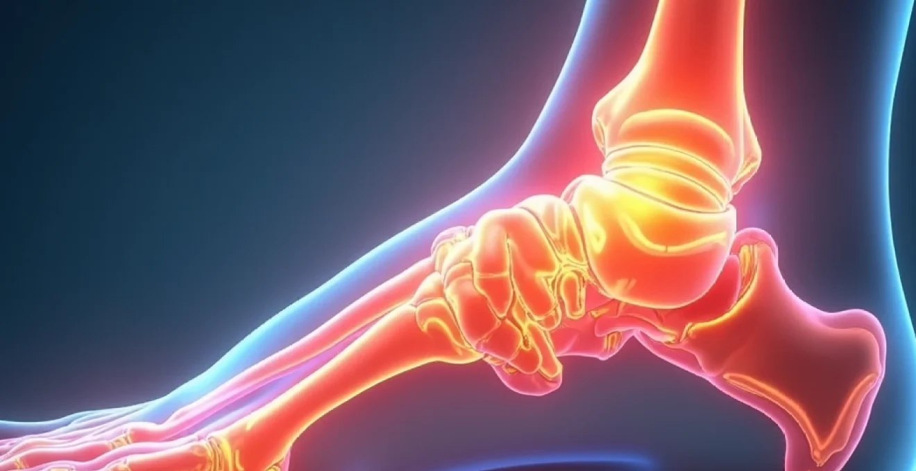
Tarsometatarsal joint stability and plantar ligament architecture
The Lisfranc joint complex encompasses the articulations between the first through fifth metatarsals and their corresponding tarsal bones, creating a sophisticated biomechanical system essential for midfoot stability. The plantar ligament architecture forms the primary stabilising structure, with the Lisfranc ligament connecting the medial cuneiform to the second metatarsal base representing the keystone of this complex. This ligament system must withstand forces exceeding three times body weight during normal gait cycles, making it particularly vulnerable to injury and subsequent surgical complications.
The intricate blood supply to this region contributes significantly to healing challenges and potential chronic pain development. Vascular compromise during surgical exposure or through direct trauma can lead to avascular necrosis, particularly affecting the second metatarsal base where blood supply is most tenuous. Surgical planning must carefully consider these anatomical constraints to minimise iatrogenic complications that contribute to long-term pain syndromes.
Open reduction internal fixation (ORIF) versus arthrodesis technique selection
Surgical technique selection significantly influences chronic pain outcomes, with both ORIF and arthrodesis presenting distinct advantages and complications profiles. ORIF procedures aim to restore anatomical alignment while preserving joint mobility, utilising various fixation methods including screws, plates, and temporary stabilisation devices. However, preserved motion can lead to ongoing mechanical stress at healing interfaces, potentially contributing to chronic inflammatory responses and persistent pain.
Primary arthrodesis eliminates joint motion but provides superior stability for severely comminuted fractures or ligamentous disruptions. While fusion procedures may resolve mechanical pain sources, they can create compensatory stress patterns in adjacent joints, leading to secondary arthritic changes and chronic discomfort. The decision between these approaches requires careful consideration of injury patterns, patient factors, and long-term functional expectations.
Kirschner wire and cannulated screw fixation methods
Hardware selection and placement techniques directly impact chronic pain development through various mechanisms including mechanical irritation, stress concentration, and biological responses. Kirschner wire fixation offers temporary stabilisation with minimal tissue disruption but requires subsequent removal procedures that can introduce additional trauma. The subcutaneous position of K-wires frequently causes skin irritation and discomfort during the healing period.
Cannulated screw fixation provides robust mechanical stability with lower profile hardware, reducing soft tissue irritation compared to plate constructs. However, screw placement accuracy becomes critical to avoid joint penetration or nerve impingement, both of which can create persistent neuropathic pain conditions. Proper surgical technique and hardware positioning remain paramount in preventing hardware-related chronic pain syndromes.
Chopart joint compensation patterns following surgical reconstruction
Midfoot surgical reconstruction inevitably alters normal biomechanical patterns, creating compensatory stress patterns that can manifest as chronic pain in adjacent joint complexes. The Chopart joint, comprising the talonavicular and calcaneocuboid articulations, frequently experiences increased loading following Lisfranc surgery as the foot adapts to altered midfoot mechanics. These compensation patterns can lead to secondary arthritic changes and chronic pain development over time.
Gait analysis studies demonstrate significant alterations in foot rollover patterns following Lisfranc reconstruction, with increased reliance on hindfoot and forefoot compensation mechanisms. Understanding these biomechanical adaptations helps clinicians anticipate potential sources of chronic pain and implement preventive measures through appropriate rehabilitation protocols and orthotic interventions.
Pathophysiology of Post-Operative lisfranc chronic pain syndrome
Complex regional pain syndrome (CRPS) development in midfoot reconstruction
Complex Regional Pain Syndrome represents one of the most challenging complications following Lisfranc surgery, affecting approximately 5-15% of patients undergoing midfoot reconstruction. CRPS development appears linked to sympathetic nervous system dysfunction triggered by surgical trauma, immobilisation, and inflammatory responses. Early recognition becomes crucial as delayed treatment significantly reduces intervention effectiveness and prolongs patient suffering.
The clinical presentation typically includes disproportionate pain, allodynia, temperature changes, and sudomotor dysfunction affecting the entire foot and potentially extending proximally.
Understanding CRPS pathophysiology requires recognising the complex interplay between peripheral nociceptive input and central sensitisation mechanisms that perpetuate pain long after tissue healing should be complete.
Early mobilisation protocols and aggressive pain management strategies can help reduce CRPS incidence, though complete prevention remains challenging.
Hardware-related pain from titanium and stainless steel implants
Implant materials and their interaction with surrounding tissues contribute significantly to chronic pain development following Lisfranc surgery. Titanium implants, while biocompatible, can create stress concentration points due to their elastic modulus differences compared to bone tissue. This mechanical mismatch can lead to micromotion at the bone-implant interface, triggering inflammatory responses and chronic pain conditions.
Stainless steel implants present additional challenges through potential corrosion and metal ion release, particularly in high-stress environments characteristic of the midfoot region. The body’s immune response to these materials can create chronic inflammatory conditions contributing to persistent pain and functional limitations. Hardware removal considerations must weigh the risks of additional surgery against potential pain relief benefits, requiring individualised patient assessment.
Arthrofibrosis and capsular contracture formation
Post-operative adhesion formation represents a significant contributor to chronic pain and functional limitation following Lisfranc surgery. The healing response naturally produces scar tissue, but excessive fibroblast activity can create restrictive adhesions that limit joint motion and create mechanical pain during attempted movement. The midfoot’s complex anatomy with multiple small joints makes it particularly susceptible to arthrofibrosis development.
Capsular contracture occurs when joint capsules become thickened and inelastic, creating pain through mechanical restriction and increased intra-articular pressure during motion attempts. Early recognition and intervention through appropriate mobilisation techniques can help minimise these complications, though established contractures often require surgical intervention for resolution.
Neuroma development in superficial peroneal and deep peroneal nerve branches
Nerve injury and subsequent neuroma formation create particularly challenging chronic pain conditions following Lisfranc surgery. The superficial peroneal nerve provides sensation to the dorsal foot and can be injured during surgical exposure or through compression from post-operative swelling and scar tissue formation. Neuromas develop when severed nerve endings attempt regeneration but encounter barriers, creating painful nodules that trigger neuropathic pain symptoms.
Deep peroneal nerve involvement can occur through direct injury or compression, creating not only sensory deficits but also motor weakness affecting toe extension. The characteristic burning, electric shock-like pain associated with neuromas requires specific treatment approaches including nerve blocks, medication management, and potentially surgical neuroma excision or nerve reconstruction procedures.
Post-traumatic arthritis progression in adjacent cuneiform joints
Arthritic changes in joints adjacent to the primary injury site represent a long-term complication that can develop months to years following initial surgery. The interconnected nature of midfoot joints means that altered mechanics from Lisfranc reconstruction creates abnormal stress patterns affecting cuneiform and navicular articulations. These secondary arthritic changes often develop insidiously, creating gradually worsening pain that may be attributed to other causes.
Cartilage degeneration occurs through both mechanical overload and inflammatory mediators released from the healing surgical site. Prevention strategies focus on restoring normal anatomical alignment and implementing early mobilisation protocols to maintain joint nutrition and prevent stiffness. However, some degree of secondary arthritis appears inevitable in many cases, requiring long-term management strategies.
Clinical assessment and diagnostic imaging for persistent lisfranc pain
Weight-bearing radiographic analysis using meary’s angle measurements
Comprehensive radiographic evaluation forms the foundation of chronic pain assessment following Lisfranc surgery, with weight-bearing studies providing crucial information about structural alignment and mechanical function. Meary’s angle measurements help identify subtle malalignment that may contribute to chronic pain through abnormal stress distribution across the midfoot complex. These angular measurements compare the longitudinal axes of the first metatarsal and talus, with normal values ranging between 0-5 degrees.
Serial radiographic monitoring allows clinicians to track progressive deformity development or hardware complications that may contribute to ongoing pain symptoms. Comparison views of the contralateral foot provide valuable reference points for assessing anatomical restoration accuracy. Advanced imaging protocols including stress views can reveal dynamic instability patterns not apparent on standard radiographs.
MRI protocol for detecting avascular necrosis in second metatarsal base
Magnetic resonance imaging provides unparalleled soft tissue detail essential for identifying complications contributing to chronic pain following Lisfranc surgery. Avascular necrosis of the second metatarsal base represents a particularly concerning complication given the tenuous blood supply to this region. MRI protocols utilising T1-weighted, T2-weighted, and STIR sequences can detect early marrow signal changes indicative of vascular compromise before radiographic changes become apparent.
The timing of MRI evaluation becomes crucial as signal changes may be influenced by normal post-operative healing responses, making interpretation challenging in the immediate post-surgical period.
Advanced MRI techniques including dynamic contrast enhancement can help differentiate between normal healing patterns and pathological vascular compromise, guiding treatment decisions for patients with persistent pain symptoms.
Bone scintigraphy SPECT-CT evaluation for occult fractures
Nuclear medicine studies offer unique insights into metabolic activity within the midfoot complex, helping identify areas of abnormal bone turnover that may contribute to chronic pain conditions. SPECT-CT fusion imaging provides both metabolic information and precise anatomical localisation, enabling detection of stress fractures, nonunions, or areas of abnormal mechanical loading not visible on conventional imaging studies.
The high sensitivity of bone scintigraphy for detecting osseous pathology makes it particularly valuable for evaluating patients with persistent pain but normal radiographic findings. Three-phase bone scans can differentiate between inflammatory conditions, infection, and mechanical problems through characteristic uptake patterns during different phases of the study.
Diagnostic lidocaine injection techniques for Joint-Specific pain localisation
Diagnostic injections represent powerful tools for pain source localisation in patients with chronic symptoms following Lisfranc surgery. Selective anaesthetic blocks of specific joints or nerve structures can help differentiate between multiple potential pain generators in the complex midfoot anatomy. These procedures require precise anatomical knowledge and fluoroscopic or ultrasound guidance for accurate needle placement.
The diagnostic value of these injections extends beyond simple pain localisation, providing information about potential treatment responses to various interventional approaches. Temporary pain relief following diagnostic blocks can predict success rates for more definitive treatments including joint injections, nerve blocks, or surgical interventions. Careful interpretation of injection results requires consideration of local anaesthetic spread patterns and patient psychology factors that may influence response patterns.
Conservative management strategies for chronic lisfranc Post-Surgical pain
Conservative management approaches form the cornerstone of chronic pain treatment following Lisfranc surgery, offering multiple therapeutic modalities that can significantly improve patient symptoms without additional surgical intervention. Multimodal pain management protocols typically combine pharmacological interventions, physical therapy, orthotic support, and injective procedures to address the complex nature of post-operative pain syndromes. The key to successful conservative management lies in identifying the primary pain mechanisms and targeting interventions accordingly.
Neuropathic pain components respond best to medications such as gabapentin, pregabalin, or tricyclic antidepressants, while inflammatory components may benefit from targeted anti-inflammatory approaches including oral medications or localised corticosteroid injections. Physical therapy protocols must be carefully designed to restore function without exacerbating pain conditions, often requiring initial pain control measures before progressing to more aggressive rehabilitation approaches. Custom orthotic devices can redistribute mechanical loads away from painful areas while supporting the altered foot architecture following surgical reconstruction.
Activity modification strategies help patients understand how to adapt daily activities to reduce pain while maintaining functional independence. This approach requires comprehensive patient education about biomechanical principles and the importance of gradual activity progression. Psychological support becomes increasingly important for patients with chronic pain conditions, as depression and anxiety frequently develop secondary to persistent symptoms and functional limitations.
Advanced conservative interventions include nerve stimulation techniques, chronic pain psychology programmes, and alternative medicine approaches such as acupuncture or massage therapy.
Success with conservative management often requires patience and persistence, as improvement may occur gradually over months rather than weeks, requiring ongoing encouragement and support from the healthcare team.
Regular reassessment allows for treatment plan modifications based on patient response and changing symptoms patterns.
The timing of conservative interventions becomes crucial, as early aggressive management often produces better outcomes than delayed treatment approaches. Some patients benefit from comprehensive pain rehabilitation programmes that address multiple aspects of chronic pain simultaneously, including physical conditioning, psychological support, medication optimisation, and functional restoration. Long-term success often depends on patient compliance with recommended treatments and realistic expectation setting regarding potential outcomes.
Revision surgery indications and advanced surgical techniques
Revision surgery consideration for chronic post-Lisfranc pain requires careful evaluation of multiple factors including pain severity, functional limitation, conservative treatment failure, and identifiable structural problems amenable to surgical correction. Hardware removal represents the most common revision procedure, particularly when implant-related pain can be clearly identified through diagnostic testing. However, hardware removal timing must consider bone healing status and the potential for instability following implant extraction.
Neurolysis procedures target nerve entrapment or neuroma formation contributing to neuropathic pain conditions. These techniques range from simple scar tissue release to complex nerve reconstruction procedures using grafting techniques. Success rates vary significantly depending on the specific nerve involved, duration of symptoms, and patient factors affecting healing capacity. Advanced microsurgical techniques have improved outcomes for complex nerve reconstruction procedures, though success cannot be guaranteed.
Corrective osteotomy procedures address malalignment issues contributing to abnormal stress patterns and chronic pain development. These procedures require precise preoperative planning using advanced imaging techniques and may involve multiple bone cuts to restore normal anatomical relationships. Computer-assisted planning and navigation systems have improved surgical accuracy for complex reconstructive procedures, though technical demands remain high.
Joint fusion procedures may be necessary for severe arthritic changes or persistent instability contributing to chronic pain conditions. Selective fusion techniques attempt to preserve motion in unaffected joints while stabilising painful articulations. The decision regarding fusion extent requires balancing pain relief benefits against functional limitations from reduced joint motion. Patient counselling regarding realistic expectations becomes crucial as revision surgery outcomes are generally less predictable than primary procedures.
Bone grafting procedures may be required for nonunion treatment or correction of established malunions contributing to chronic pain. Autograft, allograft, and synthetic bone substitute materials all have specific advantages and limitations depending on the clinical situation. Success rates for revision procedures typically decrease compared to primary surgery, making conservative management attempts increasingly important before considering surgical options.
Long-term functional outcomes and return to activity protocols
Long-term functional outcomes following Lisfranc surgery with chronic pain complications demonstrate significant variability depending on multiple factors including injury severity, surgical technique, compliance with rehabilitation protocols, and individual patient characteristics. Studies indicate that while many patients achieve acceptable pain levels and functional capacity, complete return to pre-injury activity levels occurs in fewer than 60% of cases. Understanding these outcome patterns helps set realistic expectations and guide treatment planning for patients experiencing chronic post-operative pain.
Return to athletic activities requires careful progression through supervised rehabilitation protocols that gradually increase loading and stress on the reconstructed midfoot complex. High-impact activities such as running, jumping, and cutting sports place significant demands on the Lisfranc complex and may need to be permanently modified or discontinued in some patients. Lower-impact alternatives including swimming, cycling, and walking programmes often provide acceptable fitness maintenance while reducing pain exacerbation risk.
Occupational considerations become particularly important for patients whose work involves prolonged standing, heavy lifting, or walking on
uneven surfaces. These patients may benefit from workplace accommodations or job modifications to reduce symptom exacerbation and prevent further injury. Ergonomic assessments can identify specific work demands that place excessive stress on the healing midfoot complex and suggest appropriate modifications.
Functional assessment tools including validated outcome measures help track recovery progress and identify areas requiring additional intervention. The American Orthopaedic Foot and Ankle Society (AOFAS) scoring system provides standardised measurements of pain, function, and alignment that can guide treatment decisions and monitor long-term outcomes. Patient-reported outcome measures complement objective testing by capturing the subjective impact of chronic pain on daily activities and quality of life.
Long-term follow-up protocols should extend beyond traditional healing timeframes, as complications may develop years after initial surgery. Regular monitoring allows for early detection of progressive deformity, hardware complications, or secondary arthritic changes that may require intervention. Patients should understand that Lisfranc injuries represent a lifelong condition requiring ongoing management even after successful initial treatment.
The journey from Lisfranc injury through surgical reconstruction to functional recovery represents one of orthopaedic medicine’s most challenging pathways, requiring patience, persistence, and realistic expectation setting for both patients and healthcare providers.
Successful long-term management often requires a team approach involving orthopaedic surgeons, physical therapists, pain management specialists, and sometimes mental health professionals. This multidisciplinary approach addresses the complex interplay between physical symptoms, functional limitations, and psychological adaptation that characterises chronic post-surgical pain conditions. Patient education remains crucial throughout this process, helping individuals understand their condition and actively participate in management decisions.
Future research directions focus on improving surgical techniques, developing better implant materials, and understanding genetic factors that may predict healing complications. Advanced biomechanical studies continue to refine our understanding of midfoot mechanics and guide surgical planning decisions. As our knowledge base expands, we can expect continued improvements in both treatment outcomes and our ability to prevent chronic pain development following Lisfranc surgery.