Abdominal skin conditions represent a diverse spectrum of dermatological manifestations that can significantly impact quality of life. The stomach region, encompassing the epigastric, periumbilical, and lower abdominal areas, provides a unique anatomical environment where various pathogenic organisms, allergens, and systemic conditions can manifest through distinctive cutaneous presentations. Understanding these dermatological patterns is essential for accurate diagnosis and appropriate therapeutic intervention.
The abdominal wall’s anatomy creates specific microenvironments that predispose certain areas to particular types of skin conditions. The presence of hair follicles, sebaceous glands, and areas of skin-to-skin contact or friction can influence the development and progression of various rashes. Environmental factors, personal hygiene practices, and underlying systemic conditions all contribute to the complex aetiology of abdominal skin manifestations.
Recognition of characteristic morphological features, distribution patterns, and associated symptoms enables healthcare professionals to differentiate between various conditions affecting this anatomical region. From infectious processes to autoimmune conditions, each category presents with distinctive clinical features that aid in establishing an accurate diagnosis and implementing targeted treatment strategies.
Bacterial dermatitis and infectious abdominal skin conditions
Bacterial skin infections affecting the abdominal region encompass a broad spectrum of conditions, ranging from superficial follicular involvement to deep tissue infections requiring immediate medical attention. These conditions often develop in individuals with compromised skin barriers, poor hygiene, or underlying immunocompromising conditions that facilitate bacterial colonisation and proliferation.
Cellulitis manifestations on abdominal wall and periumbilical region
Cellulitis presents as a rapidly spreading, erythematous, warm, and tender area of skin inflammation that extends into the deeper dermal and subcutaneous tissues. On the abdominal wall, cellulitis typically manifests as poorly demarcated areas of redness with associated swelling and warmth. The periumbilical region is particularly susceptible due to the presence of natural skin folds and potential for bacterial seeding through minor trauma or poor hygiene practices.
The characteristic features include expanding erythema with indistinct borders, pitting oedema, and significant tenderness upon palpation. Systemic symptoms such as fever, chills, and malaise frequently accompany severe cases, indicating the need for prompt antibiotic intervention. Streptococcus pyogenes and Staphylococcus aureus represent the most common causative organisms, with methicillin-resistant Staphylococcus aureus (MRSA) becoming increasingly prevalent in certain populations.
Impetigo lesions: Honey-Crusted vesicles and pustular formations
Impetigo on the abdominal region typically presents as superficial vesiculopustular lesions that rupture easily, leaving characteristic honey-coloured crusts. These lesions often develop following minor skin trauma, insect bites, or secondary infection of pre-existing dermatitis. The condition is highly contagious and can spread rapidly through direct contact or contaminated clothing and bedding.
Two primary forms affect the abdominal area: non-bullous impetigo, characterised by small vesicles that quickly rupture and form golden crusts, and bullous impetigo, presenting as larger fluid-filled blisters that eventually rupture, leaving collarette-like scales. The superficial nature of impetigo lesions distinguishes them from deeper bacterial infections, though secondary complications can occur if left untreated.
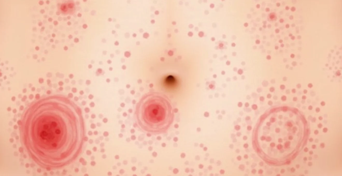
Folliculitis distribution patterns across lower torso and epigastric area
Folliculitis affecting the abdominal region demonstrates characteristic distribution patterns corresponding to hair follicle density and areas prone to friction or occlusion. The condition presents as small, erythematous papules or pustules centred around hair follicles, creating a distinctive polka-dot appearance across affected areas. Hot tub folliculitis, caused by Pseudomonas aeruginosa, may develop following exposure to contaminated water sources.
Predisposing factors include tight clothing, excessive sweating, use of comedogenic products, and compromised immune status. The lower abdominal region and areas beneath undergarments are particularly susceptible due to increased moisture retention and friction. Chronic folliculitis can lead to post-inflammatory hyperpigmentation and scarring, particularly in individuals with darker skin types.
Staphylococcal and streptococcal skin infections: clinical differentiation
Distinguishing between staphylococcal and streptococcal infections on the abdominal skin requires careful assessment of morphological characteristics and clinical progression. Staphylococcal infections typically present with well-circumscribed, purulent lesions that may develop central necrosis or abscess formation. These infections often remain localised unless host factors predispose to systemic spread.
Streptococcal infections, conversely, tend to produce rapidly spreading, poorly demarcated areas of erythema and oedema with a more inflammatory appearance. The presence of lymphangitic streaking, regional lymphadenopathy, and systemic symptoms more commonly accompanies streptococcal infections. Bacterial culture and sensitivity testing remain essential for definitive identification and appropriate antibiotic selection, particularly in cases of treatment failure or suspected antibiotic resistance.
Fungal cutaneous manifestations and dermatophyte infections
Fungal infections of the abdominal skin create distinctive clinical presentations that reflect the unique characteristics of various fungal pathogens. These conditions thrive in warm, moist environments and can persist for extended periods without appropriate antifungal intervention. Understanding the morphological variations and predisposing factors associated with different fungal species enables accurate diagnosis and targeted therapy.
Tinea corporis: annular scaling patches on abdominal surface
Tinea corporis presents on the abdominal skin as characteristic annular lesions with raised, scaly borders and relative central clearing, creating the classic “ringworm” appearance. These lesions typically begin as small, erythematous patches that gradually expand outward while the central area shows signs of healing, resulting in the pathognomonic ring-shaped configuration.
The advancing border demonstrates active inflammation with scaling, vesiculation, or pustulation, while the central area may appear normal or show mild hyperpigmentation. Multiple lesions can coalesce to form polycyclic or gyrate patterns , particularly in immunocompromised individuals or cases of delayed treatment. Trichophyton rubrum and Microsporum canis represent common causative organisms, with transmission occurring through direct contact with infected individuals, animals, or contaminated surfaces.
Candidiasis intertrigo in skin folds and inframmammary regions
Candidal intertrigo affects inframammary areas and other skin folds adjacent to the abdominal region, presenting as erythematous, macerated patches with characteristic satellite pustules at the periphery. The condition develops in areas of skin-to-skin contact where moisture accumulation and reduced air circulation create optimal conditions for Candida albicans proliferation.
The primary lesion appears as a well-demarcated, erythematous patch with a glazed or velvety surface texture. Satellite lesions consisting of small pustules or papulopustules extend beyond the main area of involvement, representing a pathognomonic feature that distinguishes candidal infections from other inflammatory conditions. Predisposing factors include obesity, diabetes mellitus, immunosuppression, and prolonged antibiotic use.
Pityriasis versicolor: hypopigmented and hyperpigmented macules
Pityriasis versicolor manifests on the abdominal skin as well-demarcated macules that may appear hypopigmented, hyperpigmented, or erythematous, depending on the individual’s baseline skin colour and sun exposure history. The condition, caused by Malassezia species, typically presents as confluent patches with fine, powdery scaling that becomes more apparent with gentle scraping.
The characteristic “spaghetti and meatballs” appearance on potassium hydroxide (KOH) preparation aids in definitive diagnosis. Lesions often become more noticeable following sun exposure, as affected areas fail to tan normally, creating a mottled appearance. The condition frequently recurs during warm, humid weather and may require maintenance antifungal therapy in susceptible individuals.
Dermatophyte identification through KOH microscopy and culture methods
Accurate identification of dermatophyte infections requires systematic laboratory evaluation combining direct microscopic examination and fungal culture techniques. KOH preparation of scale samples reveals characteristic septate hyphae and arthroconidia, confirming the presence of dermatophyte infection. However, species identification requires fungal culture on appropriate media such as Sabouraud’s dextrose agar.
Colony morphology, microscopic features, and biochemical testing enable definitive species identification, which influences treatment selection and epidemiological considerations. Dermoscopy can provide additional diagnostic clues , revealing comma hairs in tinea capitis or the characteristic scaling patterns in tinea corporis. Polymerase chain reaction (PCR) and matrix-assisted laser desorption/ionisation time-of-flight (MALDI-TOF) mass spectrometry represent emerging diagnostic modalities offering rapid and accurate species identification.
Allergic contact dermatitis and irritant reactions
Contact dermatitis affecting the abdominal region results from either allergic sensitisation or direct irritant exposure to various environmental substances. The condition presents distinct morphological patterns that often reflect the shape and distribution of the offending agent, creating characteristic geometric or linear configurations that aid in identifying potential triggers.
Allergic contact dermatitis develops through delayed-type hypersensitivity mechanisms following sensitisation to specific allergens. Common triggers affecting the abdominal area include metal components in belt buckles, clothing fasteners, or jewellery containing nickel, cobalt, or chromium. Textile allergens, including formaldehyde-releasing preservatives, dyes, and fabric finishes , can produce widespread dermatitis corresponding to areas of clothing contact.
The clinical presentation typically includes well-demarcated erythematous patches with vesiculation, weeping, and subsequent crusting in acute phases. Chronic exposure leads to lichenification, scaling, and post-inflammatory pigmentary changes. The geometric patterns often mirror the shape of belt buckles, clothing seams, or other contact surfaces, providing valuable diagnostic clues.
Irritant contact dermatitis presents similarly but develops through direct cytotoxic effects rather than immunological mechanisms. Common irritants include harsh detergents, antiseptics, adhesive tapes, and friction from tight clothing. The condition often affects individuals with compromised skin barriers , including those with atopic dermatitis or occupational exposures to irritating substances.
Patch testing remains the gold standard for identifying specific allergens in cases of allergic contact dermatitis. The procedure involves applying standardised allergen panels to the back and evaluating reactions over multiple time points. Positive reactions demonstrate characteristic morphology and timing that distinguish allergic responses from irritant reactions or false-positive results.
Understanding the distinction between allergic and irritant contact dermatitis is crucial for implementing effective avoidance strategies and preventing recurrent episodes through targeted lifestyle modifications.
Viral exanthems and systemic disease manifestations
Viral infections frequently produce characteristic cutaneous manifestations on the abdominal region as part of systemic disease processes. These exanthems often follow predictable patterns of distribution and morphology that aid in clinical diagnosis and management decisions. Understanding viral exanthem patterns enables healthcare providers to differentiate between various viral pathogens and implement appropriate isolation precautions.
Varicella-zoster distribution along dermatome T6-T12 pathways
Herpes zoster affecting thoracic dermatomes T6-T12 produces characteristic unilateral vesicular eruptions that follow specific anatomical distributions across the abdominal wall. The condition begins with prodromal symptoms including burning, tingling, or stabbing pain in the affected dermatome, followed by the development of grouped vesicles on an erythematous base.
The vesicular phase progresses through pustulation, crusting, and eventual healing over 2-4 weeks. Post-herpetic neuralgia represents a significant complication , particularly in elderly individuals or immunocompromised patients. The unilateral distribution and dermatomal pattern distinguish zoster from other vesicular conditions affecting the abdomen.
Varicella (chickenpox) presents as a generalised vesicular eruption that includes the abdominal region as part of widespread cutaneous involvement. The characteristic “dewdrop on a rose petal” appearance and presence of lesions in various stages of development (papules, vesicles, pustules, and crusts) create a distinctive clinical picture.
Hand, foot and mouth disease: coxsackievirus A16 vesicular eruptions
Hand, foot and mouth disease occasionally produces cutaneous manifestations on the abdominal region, particularly in severe cases or atypical presentations. The condition, primarily caused by Coxsackievirus A16 or Enterovirus 71, typically affects the oral mucosa, palms, soles, and occasionally extends to the trunk and abdominal areas.
Abdominal lesions appear as small vesicles or papulovesicles on an erythematous base, often accompanied by oral ulcerations and characteristic palmoplantar involvement. The distribution pattern and associated oral findings distinguish this condition from other viral exanthems. Atypical presentations may demonstrate more extensive truncal involvement, particularly in immunocompromised individuals.
Pityriasis rosea: herald patch and christmas tree distribution pattern
Pityriasis rosea frequently affects the abdominal region as part of its characteristic “Christmas tree” distribution pattern along skin tension lines. The condition begins with a herald patch, typically appearing on the trunk or proximal extremities, followed by the development of smaller secondary lesions over 1-2 weeks.
The secondary eruption demonstrates a distinctive pattern following Langer’s lines, creating the pathognomonic Christmas tree appearance when viewed from the back. Individual lesions present as oval, salmon-coloured patches with peripheral scaling and central clearing. The condition is self-limiting, typically resolving within 6-8 weeks without specific intervention, though topical therapies may provide symptomatic relief.
Molluscum contagiosum: umbilicated papules and central keratotic plugs
Molluscum contagiosum affecting the abdominal region presents as characteristic dome-shaped, flesh-coloured to pink papules with central umbilication. The lesions, caused by a poxvirus, demonstrate a waxy or pearlescent quality and may develop central keratotic plugs that can be expressed with gentle pressure.
The condition spreads through direct contact or auto-inoculation, leading to clustered distributions in areas of trauma or scratching. Immunocompromised individuals may develop numerous, larger lesions that persist for extended periods. The characteristic morphology and central dell distinguish molluscum from other papular conditions affecting the abdominal skin.
Autoimmune and inflammatory dermatological conditions
Autoimmune and chronic inflammatory conditions affecting the abdominal skin present complex clinical challenges requiring comprehensive diagnostic evaluation and long-term management strategies. These conditions often demonstrate characteristic morphological features and distribution patterns that aid in differential diagnosis, though definitive identification frequently requires histopathological examination and immunofluorescence studies.
Psoriasis commonly affects the abdominal region, presenting as well-demarcated, erythematous plaques with characteristic silvery scaling. The lesions demonstrate the pathognomonic Auspitz sign when scales are removed, revealing punctate bleeding points. Koebner phenomenon may lead to linear lesions following trauma or surgical incisions across the abdominal wall. The condition often demonstrates symmetrical distribution and may be associated with nail changes, arthritis, and metabolic comorbidities.
Eczematous dermatitis affecting the abdominal region encompasses various subtypes including atopic dermatitis, nummular eczema, and seborrhoeic dermatitis. Atopic dermatitis typically presents as ill-defined, erythematous patches with excoriation and lichenification in chronic cases. The condition often demonstrates
a predilection for flexural areas and may be accompanied by intense pruritus that worsens during nighttime hours.Lichen planus occasionally manifests on the abdominal skin as violaceous, polygonal papules with characteristic Wickham’s striae visible under dermoscopy. The condition may present as isolated lesions or as part of generalised cutaneous involvement. *Oral mucosal involvement frequently accompanies cutaneous manifestations*, requiring comprehensive examination of all potentially affected sites.Systemic lupus erythematosus can produce various cutaneous manifestations on the abdominal region, including non-specific erythematous patches, discoid lesions, or photosensitive eruptions. The characteristic malar rash may extend to include the upper chest and abdominal areas in severe cases. Laboratory evaluation including antinuclear antibodies, anti-double-stranded DNA, and complement levels aids in establishing the diagnosis and monitoring disease activity.Dermatomyositis presents with distinctive cutaneous features that may extend to the abdominal region, including erythematous to violaceous patches with associated muscle weakness and elevated muscle enzymes. *The pathognomonic heliotrope rash and Gottron’s papules* help distinguish this condition from other autoimmune dermatoses. Malignancy screening is essential in adult-onset cases due to the increased risk of underlying neoplasms.
Parasitic infestations and arthropod-related skin reactions
Parasitic infestations affecting the abdominal skin create distinctive clinical presentations that require prompt recognition and appropriate treatment to prevent complications and transmission to close contacts. These conditions often demonstrate characteristic distribution patterns and morphological features that aid in differential diagnosis, though definitive identification may require specialised laboratory techniques.Scabies infestation commonly affects the abdominal region, presenting as intensely pruritic papules, vesicles, and characteristic burrows in skin folds and areas of thin skin. The pathognomonic burrows appear as short, serpentine tracks with a vesicle or papule at one end, representing the location of the gravid female mite. *Nocturnal intensification of pruritus* represents a hallmark feature that distinguishes scabies from other pruritic conditions.The distribution pattern typically involves the periumbilical area, lower abdominal region, and areas beneath undergarments. Secondary bacterial infection may develop due to excoriation and compromised skin integrity. Norwegian or crusted scabies may occur in immunocompromised individuals, presenting as hyperkeratotic plaques with millions of mites, creating a highly contagious condition requiring isolation precautions.Pediculosis corporis, though less common than head or pubic lice infestations, can affect the abdominal region in individuals with poor hygiene or overcrowded living conditions. The condition presents as erythematous papules and excoriation marks in areas of clothing contact. *Adult lice and nits may be identified in clothing seams* rather than directly on the skin, distinguishing this condition from other ectoparasitic infestations.Arthropod bites and stings affecting the abdominal region demonstrate varied morphology depending on the specific arthropod involved and individual host responses. Flea bites typically present as small, erythematous papules with central puncta, often arranged in linear or clustered patterns corresponding to areas of pet contact or infested environments. The lesions may develop vesiculation or bullous changes in sensitive individuals.Bed bug bites characteristically appear as linear arrangements of three or more erythematous papules, often referred to as “breakfast, lunch, and dinner” lesions. The abdominal region frequently demonstrates these characteristic patterns, particularly in areas exposed during sleep. *Delayed hypersensitivity reactions may develop*, leading to persistent, intensely pruritic lesions that can become secondarily infected.Spider bites require careful evaluation to distinguish between harmless species and potentially dangerous arachnids such as black widow or brown recluse spiders. Most spider bites present as localised erythematous papules or plaques with minimal systemic symptoms. However, brown recluse envenomation can produce progressive tissue necrosis with characteristic “bull’s-eye” appearance and central eschars.Tick bites may occur on the abdominal region during outdoor activities, presenting as erythematous papules or plaques at attachment sites. The development of erythema migrans, characterised by expanding annular erythema with central clearing, indicates Lyme disease and requires prompt antibiotic intervention. *Embedded tick parts may persist after removal*, leading to persistent inflammatory reactions or secondary bacterial infection.Chigger bites typically affect areas where clothing fits tightly against the skin, including the waistline and lower abdominal region. The lesions present as intensely pruritic, erythematous papules or papulovesicles that may persist for several weeks. The microscopic nature of chigger larvae makes visual identification difficult, requiring clinical correlation with exposure history and characteristic distribution patterns.Cutaneous larva migrans, caused by penetration of animal hookworm larvae through the skin, creates pathognomonic serpentine tracks that advance daily as the larvae migrate through the epidermis. The abdominal region may be affected following contact with contaminated sand or soil, particularly in tropical or subtropical environments. *The advancing edge of the track often demonstrates active inflammation and vesiculation*, while older portions may show resolution with post-inflammatory changes.Human botfly myiasis represents an uncommon but distinctive parasitic infestation that may affect the abdominal skin following travel to endemic areas. The condition presents as nodular lesions with central puncta that may demonstrate periodic movement or discharge. The larvae require surgical extraction, as topical treatments are generally ineffective for established infections.Treatment of parasitic infestations requires identification of the specific organism and implementation of appropriate antiparasitic therapy. Scabies responds to topical permethrin or oral ivermectin, with treatment of all household contacts and environmental decontamination essential for preventing reinfestation. *Contact tracing and treatment of close contacts* represents a crucial component of successful parasite control, particularly in institutional settings or among vulnerable populations.