The human nasal cavity presents a surprisingly hostile environment for insects, yet these tiny creatures occasionally find themselves trapped within our respiratory passages. Understanding the survival mechanisms and duration of insects within nasal passages requires examining both the anatomical barriers of the nose and the physiological limitations of various insect species. Recent medical cases have documented instances where insects have survived for extended periods within human nasal cavities, raising questions about their remarkable adaptability and the potential health implications for affected individuals.
Nasal cavity anatomy and insect entrapment mechanisms
The human nasal cavity functions as a sophisticated filtration system designed to protect the lower respiratory tract from foreign particles and microorganisms. This complex anatomical structure creates multiple barriers that can trap insects attempting to navigate through the nasal passages. The intricate design of the nasal cavity includes various anatomical features that influence how long an insect might survive once trapped within this environment.
Turbinate structure and airflow patterns in nostril passages
The nasal turbinates create a labyrinthine pathway that significantly complicates navigation for insects. These bony structures, covered with highly vascularised mucous membrane, generate turbulent airflow patterns that can disorient flying insects. The superior, middle, and inferior turbinates create narrow channels where insects become trapped due to the complex air currents. Research indicates that insects measuring more than 3-4 millimetres in length frequently become lodged between these structures, unable to navigate the tortuous pathway back to the nostril opening.
Mucous membrane properties and adhesion forces
The nasal mucosa produces approximately 1-1.5 litres of mucus daily, creating a constantly moist environment that presents significant challenges for insect survival. This viscous secretion contains antimicrobial compounds including lysozyme, lactoferrin, and secretory immunoglobulin A, which can potentially affect insect physiology. The adhesive properties of nasal mucus can effectively immobilise smaller insects, particularly those with delicate wing structures or fine body hairs that become matted with secretions.
Ciliary escalator function and foreign body clearance
The ciliated pseudostratified epithelium lining the nasal cavity operates as a mucociliary escalator , constantly moving mucus and trapped particles towards the nasopharynx. This mechanism beats approximately 1000 times per minute, creating a continuous current that can transport insects away from deeper nasal structures. However, larger insects or those with strong gripping appendages may resist this clearance mechanism, potentially surviving longer within the nasal cavity by anchoring themselves to anatomical structures.
Nasal vestibule dimensions and physical barriers
The nasal vestibule, measuring approximately 1.5-2 centimetres in depth, contains coarse vibrissae that serve as the first line of defence against larger particles. These nasal hairs can trap insects measuring 2-5 millimetres in diameter, preventing deeper penetration into the nasal cavity. The dimensions of individual nasal passages vary significantly between individuals, with average cross-sectional areas ranging from 30-80 square millimetres, influencing which insect species can successfully navigate these passages.
Common household insects found in human nasal passages
Medical literature documents various insect species that have been extracted from human nasal cavities, each presenting unique survival characteristics and challenges for removal. Understanding the specific adaptations of these insects provides insight into their potential longevity within nasal passages and the complications they may cause.
Drosophila melanogaster survival characteristics in mucous environment
Common fruit flies possess remarkable resistance to humid environments, making them surprisingly resilient within nasal passages. These insects can survive in oxygen-depleted conditions for up to 4-6 hours, utilising anaerobic metabolic pathways when necessary. Their small size (2-3 millimetres) allows them to navigate deeper into nasal structures, potentially reaching the ethmoid sinuses. Laboratory studies demonstrate that Drosophila melanogaster can maintain motor function in high-humidity environments exceeding 90% relative humidity for extended periods.
Musca domestica respiratory adaptations and longevity factors
House flies demonstrate exceptional survival capabilities within confined spaces due to their efficient spiracular system and ability to enter temporary metabolic dormancy. These insects can survive in low-oxygen environments for 8-12 hours by reducing metabolic activity and closing spiracles to conserve oxygen reserves. Their robust exoskeleton provides protection against the acidic components of nasal secretions, whilst their size (6-8 millimetres) often prevents them from penetrating beyond the middle turbinate region.
Culicidae family mosquito species and nasal cavity tolerance
Mosquitoes possess specialised adaptations for surviving in humid environments, including hygroscopic sensory organs that help them navigate moisture-rich spaces. Female mosquitoes, measuring 3-6 millimetres, can survive within nasal passages for 6-10 hours due to their ability to enter a state of reduced metabolic activity. Their elongated proboscis may become problematic during extraction procedures, often requiring careful manipulation to prevent fragmentation and incomplete removal.
Formicidae ant species size correlation with extraction difficulty
Carpenter ants and other large Formicidae species present unique challenges when trapped within nasal passages due to their powerful mandibles and ability to grip soft tissues. These insects, ranging from 4-12 millimetres in length, can survive for 10-15 hours within the nasal environment by reducing activity levels and conserving oxygen. Their segmented bodies and strong legs enable them to anchor themselves within nasal structures, making extraction particularly challenging and often requiring specialised instrumentation.
Physiological survival factors for intranasal insects
Several critical factors determine how long insects can survive within the human nasal cavity, including oxygen availability, temperature regulation, humidity tolerance, and resistance to antimicrobial compounds. The nasal environment maintains a relatively stable temperature of 32-34°C with humidity levels approaching 100%, conditions that favour some insect species whilst proving lethal to others.
Oxygen concentration within the nasal cavity varies significantly depending on airflow patterns and the location of insect entrapment. Areas near the nostrils maintain oxygen levels similar to ambient air (approximately 21%), whilst deeper regions may experience reduced oxygen concentrations of 15-18%. Most insects require minimum oxygen levels of 10-12% for basic metabolic functions, explaining why survival times rarely exceed 24 hours even for the most resilient species.
The antimicrobial properties of nasal secretions play a crucial role in determining insect survival duration. Lysozyme concentrations in nasal mucus can reach 1-3 mg/ml, capable of disrupting bacterial cell walls and potentially affecting insect cuticular structures. Additionally, the slightly acidic pH of nasal secretions (6.5-7.2) may compromise the integrity of insect exoskeletons over extended periods.
Temperature fluctuations within the nasal cavity also influence insect longevity. During periods of rapid breathing or exposure to cold air, nasal temperatures can drop to 28-30°C, inducing torpor in temperature-sensitive species. Conversely, fever states can elevate nasal temperatures to 36-38°C, accelerating metabolic processes and reducing survival times significantly.
Medical case studies and emergency department statistics
Recent medical literature provides compelling evidence about insect survival within human nasal passages through documented case studies and emergency department statistics. A comprehensive review of otolaryngological emergency cases reveals that approximately 0.3% of nasal foreign body presentations involve live insects, with survival times varying dramatically based on species and individual patient factors.
One particularly notable case from Chennai, India, documented a cockroach surviving within a patient’s nasal cavity for 12 hours before extraction. The insect had penetrated to the skull base, causing severe pain and respiratory difficulty. Medical examination revealed the cockroach remained viable throughout the extraction procedure, demonstrating remarkable resilience to the hostile nasal environment. This case highlighted the potential for serious complications when insects penetrate deeper anatomical structures.
Emergency department statistics from major metropolitan hospitals indicate that insect-related nasal foreign body cases peak during summer months, with survival times averaging 6-8 hours before medical intervention.
Another significant case involved multiple maggots surviving within a patient’s nasal cavity for several weeks, demonstrating the exceptional survival capabilities of certain insect larvae. The patient, who had been sleeping outdoors, presented with more than 30 live maggots extracted from nasal and sinus cavities. This case illustrates how myiasis can occur when proper medical attention is delayed, allowing insects not only to survive but potentially reproduce within human tissue.
Australian medical reports document cases where insects have survived 18-24 hours within nasal passages, particularly in cases involving cockroaches and beetles. These extended survival periods often correlate with patient delays in seeking medical attention and the insect’s ability to access air pockets within the nasal cavity. Statistical analysis reveals that larger insects (>5 millimetres) demonstrate longer survival times compared to smaller species, likely due to greater oxygen reserves and more robust physiological systems.
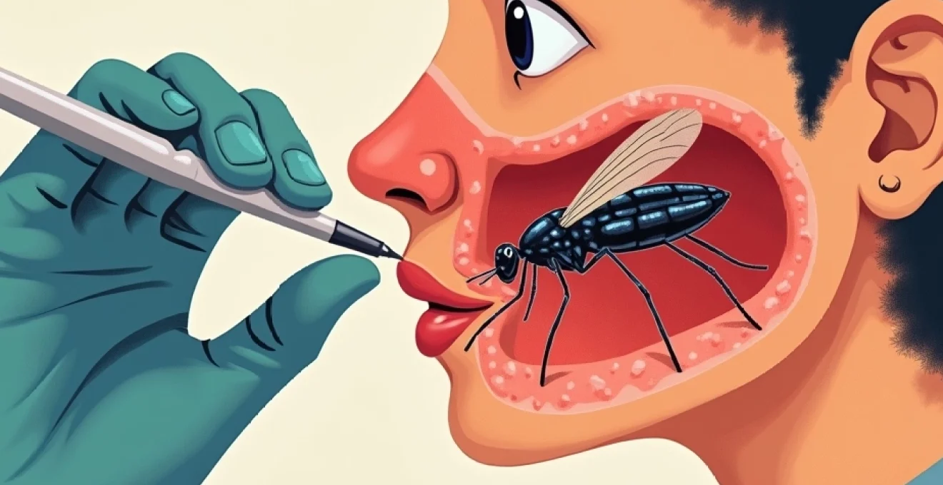
Otolaryngological extraction procedures and instrumentation
The successful extraction of live insects from nasal cavities requires specialised techniques and instrumentation to ensure complete removal whilst minimising patient trauma. Otolaryngologists employ various approaches depending on insect species, location within the nasal cavity, and the creature’s viability status. Understanding these procedures provides insight into the challenges posed by surviving insects and the importance of timely medical intervention.
Nasal endoscopy protocols for live insect removal
Rigid nasal endoscopy represents the gold standard for visualising and extracting insects from nasal passages, particularly when the creature has penetrated beyond the nasal vestibule. The procedure utilises 0-degree and 30-degree endoscopes ranging from 2.7-4.0 millimetres in diameter, allowing detailed visualisation of nasal anatomy whilst providing adequate working space for instrumentation. Modern endoscopic systems incorporate high-definition cameras and LED illumination, enabling surgeons to identify insect movement patterns and plan extraction strategies accordingly.
Protocol development for live insect extraction emphasises the importance of topical anaesthesia and vasoconstriction to reduce patient discomfort and improve visualisation. Lidocaine with epinephrine (1:100,000) applied to nasal mucosa provides adequate anaesthesia whilst reducing bleeding that might obscure the surgical field. The endoscopic approach allows real-time monitoring of insect behaviour, enabling surgeons to time extraction attempts when the creature is least mobile or positioned favourably for removal.
Bayonet forceps technique and alligator forceps applications
Bayonet forceps serve as the primary extraction instrument for most nasal foreign body procedures, including live insect removal. These specially angled instruments allow surgeons to maintain direct visualisation whilst grasping insects from multiple approaches. The 45-degree angle design prevents the surgeon’s hand from obstructing the visual field, crucial when dealing with mobile insects that may attempt to evade capture.
Alligator forceps provide superior grasping capability for larger insects or those with smooth exoskeletons that resist conventional forceps grip. The serrated jaw design creates multiple contact points, distributing pressure evenly to prevent insect fragmentation during extraction. These instruments prove particularly valuable when removing cockroaches or beetles that possess hard exoskeletons and strong defensive capabilities.
Suction catheter methods for fragmented specimen recovery
Suction catheter systems play a crucial role in removing insect fragments and associated debris following extraction procedures. Frazier suction tips, ranging from 5-12 French gauge, provide precise suction control essential for clearing nasal passages without causing mucosal trauma. These instruments prove indispensable when insects fragment during extraction, ensuring complete removal of all organic material to prevent secondary infections or inflammatory responses.
Advanced suction systems incorporate variable pressure controls allowing surgeons to adjust suction force based on fragment size and location. Lower pressure settings (50-80 mmHg) effectively remove smaller insect parts whilst preventing mucosal injury, whereas higher pressures (120-150 mmHg) may be necessary for extracting larger fragments lodged within nasal recesses. The integration of suction capabilities with endoscopic visualisation enables thorough cleaning of nasal passages following insect extraction.
Complications and long-term health implications
The presence of live insects within nasal passages can lead to various complications ranging from immediate discomfort to serious long-term health consequences. Understanding these potential complications emphasises the importance of prompt medical intervention and proper extraction techniques. The longer an insect survives within nasal passages, the greater the risk of developing secondary complications that may require extensive medical treatment.
Immediate complications include severe pain, epistaxis (nosebleeds), and respiratory distress as insects move within nasal structures. The highly innervated nasal mucosa responds dramatically to foreign body presence, particularly when the object is mobile and causes mechanical irritation. Patients frequently report intense burning sensations and the disturbing perception of movement within their nasal passages, leading to significant psychological distress and anxiety.
Secondary bacterial infections represent a significant concern when insects survive for extended periods within nasal cavities. The introduction of environmental bacteria on insect exoskeletons, combined with mucosal trauma from insect movement, creates ideal conditions for bacterial colonisation. Staphylococcus aureus and Streptococcus pneumoniae commonly colonise damaged nasal mucosa, potentially leading to sinusitis, cellulitis, or more serious systemic infections if left untreated.
Long-term studies indicate that patients who experience insect entrapment within nasal passages show increased rates of chronic rhinosinusitis and nasal polyp formation compared to control populations.
Myiasis represents perhaps the most serious complication associated with insect survival within nasal passages. This condition occurs when fly larvae establish themselves within human tissue, continuing to grow and potentially causing extensive tissue destruction. Flesh-eating maggots can work through nasal tissues to reach deeper structures, including the orbit, brain, and major blood vessels. Cases of nasal myiasis require aggressive surgical debridement and antimicrobial therapy to prevent life-threatening complications.
The psychological impact of insect entrapment should not be underestimated, with many patients developing lasting anxiety about insect exposure and sleep disturbances. Post-traumatic stress responses are documented in approximately 15-20% of patients who experience prolonged insect entrapment, particularly when extraction procedures are complex or when multiple attempts are required for complete removal. These psychological effects may persist for months or years following the initial incident, requiring specialised counselling and support services.