Kenalog injection dents represent one of the most distressing complications following corticosteroid therapy, affecting patients who receive these treatments for various dermatological and inflammatory conditions. These localised depressions in the skin, medically termed steroid-induced lipoatrophy, occur when triamcinolone acetonide disrupts the normal structure of subcutaneous fat and dermal collagen. The psychological impact of these visible indentations often matches or exceeds the physical discomfort, particularly when they appear on exposed areas such as the face, scalp, or arms. Understanding the mechanisms behind these complications and the comprehensive treatment options available provides hope for patients experiencing this challenging side effect.
Understanding kenalog injection site atrophy and subcutaneous fat necrosis
Steroid-induced atrophy develops through a complex cascade of cellular disruptions that fundamentally alter the architecture of dermal and subcutaneous tissues. When triamcinolone acetonide crystals deposit in tissues, they create localised areas of intense anti-inflammatory activity that inadvertently damage healthy structures. The corticosteroid’s mechanism involves binding to glucocorticoid receptors within fibroblasts, effectively shutting down collagen synthesis whilst simultaneously increasing collagenase activity. This dual action creates an imbalance where tissue breakdown exceeds regeneration, leading to the characteristic depressed appearance of injection sites.
Triamcinolone acetonide mechanism and dermal impact assessment
The molecular impact of triamcinolone acetonide extends beyond simple anti-inflammatory effects, fundamentally altering cellular metabolism in the injection zone. Corticosteroids suppress fibroblast proliferation by interfering with growth factor signalling pathways, particularly those involving transforming growth factor-beta and platelet-derived growth factor. Additionally, the medication disrupts normal angiogenesis, reducing blood flow to the affected area and compromising nutrient delivery essential for tissue maintenance. These changes create a localised environment where existing collagen fibres deteriorate whilst new synthesis remains severely impaired.
Identifying Steroid-Induced lipoatrophy vs normal Post-Injection changes
Distinguishing between temporary post-injection swelling and permanent atrophic changes requires careful clinical observation over time. Normal injection site reactions typically resolve within 48-72 hours, presenting as mild erythema and slight swelling that gradually subsides. In contrast, steroid-induced atrophy manifests as a progressive depression that becomes more pronounced over weeks to months following the injection. The affected area often displays characteristic features including skin thinning, visible capillary networks beneath the surface, and a distinctive shiny or stretched appearance that differs markedly from surrounding healthy tissue.
Clinical presentation of Corticosteroid-Related skin depression
The clinical presentation of Kenalog-induced depressions varies significantly depending on injection depth, concentration, and individual patient factors. Superficial injections typically produce shallow, well-demarcated depressions with minimal surrounding tissue changes, whilst deeper administrations can create more extensive atrophy affecting both dermal and subcutaneous layers. Patients frequently report a gradual onset of the depression, often noting that the area initially appeared normal before developing the characteristic sunken appearance over several weeks. The depressions may also exhibit altered pigmentation, ranging from hypopigmentation in darker skin tones to a translucent quality in lighter complexions.
Histological changes in adipose tissue following intralesional steroid administration
Microscopic examination of steroid-affected tissues reveals profound structural alterations that explain the visible clinical changes. Adipocytes undergo significant size reduction and eventual cell death through a process involving disrupted lipid metabolism and increased lipolysis. The extracellular matrix shows marked collagen fragmentation with reduced cross-linking between fibres, creating a weakened scaffold that cannot maintain normal tissue architecture. Vascular changes include endothelial cell damage and reduced capillary density, further compromising the tissue’s ability to heal and regenerate. These histological findings underscore why recovery requires extended periods and often benefits from active therapeutic intervention.
Non-invasive treatment protocols for Kenalog-Induced skin depressions
Non-invasive treatment approaches offer the first line of intervention for patients with mild to moderate steroid-induced atrophy, providing safer alternatives to surgical procedures whilst promoting natural tissue regeneration. These protocols focus on stimulating collagen production, improving local circulation, and supporting the skin’s natural healing processes through topical agents and energy-based treatments. The success of non-invasive methods depends heavily on early intervention, with treatments initiated within the first six months post-injection showing significantly better outcomes than delayed therapeutic approaches.
Topical retinoid therapy using tretinoin and adapalene applications
Retinoid therapy represents a cornerstone of non-invasive treatment, leveraging these compounds’ proven ability to stimulate collagen synthesis and accelerate cellular turnover. Tretinoin cream in concentrations ranging from 0.025% to 0.1% demonstrates particular efficacy when applied consistently over extended periods. The mechanism involves binding to retinoic acid receptors in dermal fibroblasts, upregulating genes responsible for collagen I and III production whilst simultaneously reducing matrix metalloproteinase activity. Clinical protocols typically begin with lower concentrations applied every other evening, gradually increasing frequency as tolerance develops. Adapalene gel offers an alternative with better tolerability profiles, particularly suitable for patients with sensitive skin or those experiencing retinoid-induced irritation.
Dermal massage techniques with vitamin E and Collagen-Stimulating serums
Structured massage protocols incorporating specific topical agents can significantly enhance tissue regeneration in atrophic areas. The mechanical stimulation of massage increases local blood flow whilst potentially breaking down fibrous adhesions that contribute to the depressed appearance. Vitamin E oil, applied during massage sessions, provides antioxidant protection whilst supporting membrane stability in regenerating cells. Professional techniques involve circular motions with gradually increasing pressure, performed for 10-15 minutes twice daily. Collagen-stimulating serums containing peptides, growth factors, or amino acid complexes amplify the benefits when applied immediately following massage sessions, maximising penetration into the prepared tissue.
LED phototherapy and Low-Level laser treatment protocols
Light-based therapies utilise specific wavelengths to stimulate cellular processes essential for tissue repair and regeneration. Red LED therapy at 660-670 nanometres demonstrates particular efficacy in promoting fibroblast activity and increasing adenosine triphosphate production within target cells. Treatment protocols typically involve 20-30 minute sessions performed 2-3 times weekly, with visible improvements often apparent after 8-12 weeks of consistent therapy. Low-level laser therapy using similar wavelengths provides more concentrated energy delivery, potentially accelerating treatment timelines whilst requiring professional administration. These modalities work synergistically with topical treatments, creating an optimal environment for tissue repair.
Microneedling with growth factor infusion for tissue regeneration
Microneedling therapy creates controlled micro-injuries that trigger the body’s natural healing response whilst simultaneously facilitating deep penetration of therapeutic agents. Professional treatments using needle depths of 1.5-2.5 millimetres prove most effective for addressing dermal atrophy, stimulating new collagen formation in the deeper layers where steroid damage typically occurs. The procedure becomes significantly more effective when combined with growth factor serums or platelet-rich plasma, which flood the created microchannels with regenerative compounds. Treatment schedules typically involve sessions every 4-6 weeks, with most patients requiring 3-6 treatments to achieve optimal results. The inflammatory cascade initiated by microneedling essentially reverses the anti-inflammatory damage caused by the original steroid injection.
Advanced dermatological interventions for severe atrophic scarring
When non-invasive treatments prove insufficient for addressing severe steroid-induced atrophy, advanced dermatological interventions offer more aggressive approaches with higher success rates. These procedures require specialist expertise and carry increased risks compared to conservative treatments, but they provide definitive solutions for cases where natural regeneration has plateaued. The decision to pursue advanced interventions typically occurs after 6-12 months of conservative treatment without satisfactory improvement, particularly in cases where the atrophy significantly impacts quality of life or professional function.
Hyaluronic acid filler injection with juvederm and restylane products
Dermal fillers provide immediate volume correction for steroid-induced depressions whilst offering additional regenerative benefits through their interaction with surrounding tissues. Hyaluronic acid-based products demonstrate particular suitability due to their biocompatibility and gradual absorption characteristics that allow for tissue adaptation over time. Juvederm Ultra and Restylane Lyft represent optimal choices for deeper depressions, whilst Restylane Silk proves more appropriate for superficial atrophic areas requiring subtle correction. The injection technique requires careful attention to depth and distribution, often utilising a tunneling approach to achieve even volume distribution without creating additional tissue trauma. Results typically last 12-18 months, during which time the hyaluronic acid’s hydrophilic properties support improved tissue hydration and potentially stimulate endogenous collagen production.
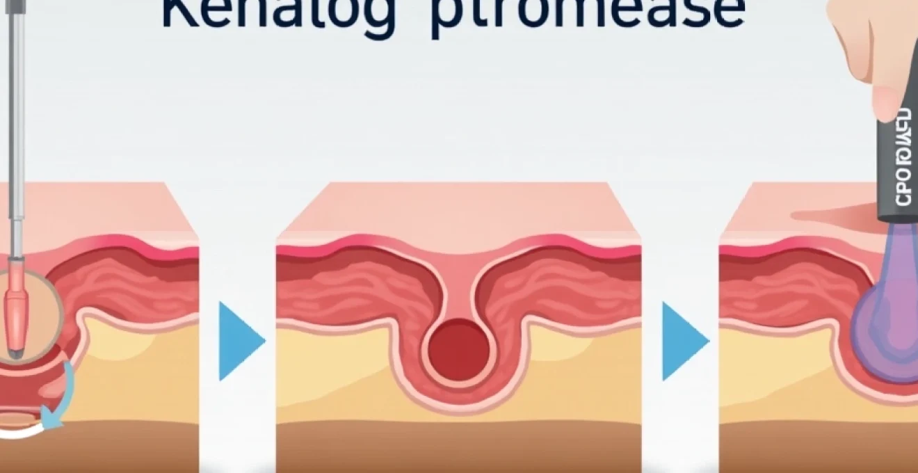
Subcision technique for fibrous band release and tissue mobilisation
Subcision addresses the underlying architectural problems in steroid-induced atrophy by mechanically releasing fibrous adhesions and creating space for new tissue formation. The procedure involves inserting a specialised needle beneath the depressed area and performing controlled cutting motions to sever restrictive bands whilst simultaneously stimulating bleeding that promotes healing responses. This technique proves particularly effective for depressions with well-defined borders and significant depth variation compared to surrounding tissue. The controlled trauma initiates inflammatory cascades that directly counteract the anti-inflammatory damage from the original steroid injection. Recovery typically requires 7-14 days, during which controlled inflammation gradually fills the released space with new connective tissue.
Fractional CO2 laser resurfacing and ablative treatment parameters
Fractional carbon dioxide laser therapy delivers controlled thermal energy to create precise zones of tissue ablation that stimulate comprehensive dermal remodelling. The treatment parameters for steroid-induced atrophy typically involve moderate energy settings with high density coverage to maximise collagen stimulation whilst maintaining safety margins. Professional protocols often utilise energy levels between 30-60 millijoules with coverage densities of 15-25%, creating thousands of microscopic treatment zones that trigger intensive healing responses. The fractional approach preserves islands of healthy tissue between treatment zones, accelerating recovery whilst minimising complications. Multiple sessions spaced 6-8 weeks apart typically achieve optimal results, with improvements continuing for 6-12 months following the final treatment as new collagen matures.
Poly-l-lactic acid stimulation using sculptra for Long-Term volume restoration
Sculptra injections offer a unique approach to treating steroid-induced atrophy by stimulating gradual, long-lasting collagen production rather than providing immediate volume correction. The poly-L-lactic acid particles act as a scaffold for new collagen formation, creating sustainable improvements that can last 2-3 years following treatment completion. The treatment protocol typically involves a series of 2-3 injection sessions spaced 4-6 weeks apart, with results becoming apparent 6-8 weeks after each treatment as new collagen develops. The gradual nature of improvement allows for natural tissue integration and reduces the risk of overcorrection that can occur with immediate-effect fillers. This approach proves particularly valuable for larger areas of atrophy or cases where multiple injection sites require treatment.
Autologous fat transfer and regenerative medicine approaches
Autologous fat transfer represents the most comprehensive solution for extensive steroid-induced atrophy, providing both immediate volume correction and long-term regenerative benefits through stem cell activity within transferred adipose tissue. This approach proves particularly valuable for cases involving large areas of atrophy or situations where multiple injection sites require simultaneous correction. The procedure involves harvesting fat from donor sites using gentle liposuction techniques, processing the adipose tissue to concentrate viable cells, and strategically injecting the prepared fat into the atrophic areas. Success rates vary significantly based on injection technique, with survival rates of 30-60% considered typical for facial applications and slightly higher rates achievable in areas with better vascularisation.
The regenerative medicine aspect of fat transfer extends beyond simple volume replacement, as transferred adipose tissue contains multipotent stem cells that can differentiate into various cell types needed for tissue repair. These adipose-derived stem cells secrete growth factors and cytokines that promote angiogenesis, stimulate resident fibroblast activity, and create an environment conducive to tissue regeneration. Recent advances in fat processing techniques, including mechanical emulsification and enzyme treatment, have improved the viability and integration of transferred cells. The procedure typically requires local anaesthesia for donor site harvesting and injection, with recovery periods of 1-2 weeks for return to normal activities. Multiple treatment sessions may be necessary to achieve optimal correction, particularly for severe cases or areas with compromised vascularisation.
Platelet-rich plasma therapy often accompanies fat transfer procedures to enhance outcomes through concentrated growth factor delivery. The combination of stem cell-rich adipose tissue with platelet-derived healing factors creates a synergistic effect that maximises regenerative potential. This combined approach has shown particular promise for treating extensive atrophy resulting from multiple injection sites or high-concentration steroid exposures. Long-term studies indicate that successful fat transfer can provide permanent correction with natural aging characteristics, making it an attractive option for younger patients seeking definitive treatment solutions.
Clinical studies demonstrate that fat grafting provides simple, effective and safe correction of corticosteroid-induced cutaneous atrophy with very satisfying aesthetic and functional results, particularly when other treatment modalities have proven insufficient.
Prevention strategies and injection technique optimisation
Preventing steroid-induced atrophy requires careful consideration of multiple factors including injection technique, medication concentration, anatomical location, and patient-specific risk factors. Healthcare providers can significantly reduce complication rates through proper training in injection methodology and adherence to established safety protocols. The concentration of triamcinolone acetonide plays a crucial role in atrophy risk, with studies showing a 3.3% incidence at 5mg/mL concentrations compared to 20% at 10mg/mL. These statistics underscore the importance of using the lowest effective concentration for achieving therapeutic goals whilst minimising adverse effects.
Injection depth represents another critical variable, with superficial injections into the dermal layer carrying higher risks of visible atrophy compared to deeper subcutaneous or intra-articular administrations. Proper needle selection and insertion angle help ensure appropriate depth placement, whilst slow injection rates allow for better tissue accommodation and reduced pressure trauma. The injection volume should be distributed across multiple sites rather than concentrated in single locations, reducing the local concentration of steroid crystals that contribute to tissue damage. Post-injection massage, when appropriate, can help distribute the medication more evenly and potentially reduce crystallisation in localised areas.
Patient education forms an essential component of prevention strategies, ensuring individuals understand both the benefits and potential risks associated with steroid injections. Those with previous adverse reactions to corticosteroids, thin skin, or compromised healing responses may require alternative treatment approaches or enhanced monitoring protocols. Documentation of injection sites, concentrations, and techniques allows for better tracking of patient responses and identification of emerging complications before they become severe. Regular follow-up appointments enable early intervention if atrophic changes begin to develop, potentially limiting the extent of tissue damage through prompt treatment modification.
Recovery timeline expectations and Long-Term prognosis assessment
Understanding realistic recovery timelines proves essential for managing patient expectations and optimising treatment outcomes following steroid-induced atrophy. Natural recovery without intervention typically occurs over 2-6 months for mild cases, though some patients may require up to 12-18 months for complete resolution. The recovery process follows a predictable pattern, beginning with cessation of progressive deepening around 4-6 weeks post-injection, followed by gradual tissue regeneration that becomes noticeable after 8-12 weeks. However, these timelines assume no additional steroid exposure to the affected area and optimal conditions for tissue healing.
Several factors influence recovery speed and completeness, including the patient’s age, overall health status, smoking history, and concurrent medications that may affect healing. Younger patients typically demonstrate faster and more complete recovery due to enhanced cellular regenerative capacity and better vascularisation. Conversely, individuals with diabetes, autoimmune conditions, or those taking medications that impair wound healing may experience prolonged recovery periods or incomplete resolution. The original injection concentration and volume also significantly impact recovery potential, with higher concentrations and larger volumes generally requiring longer healing periods.
Long-term prognosis for treated steroid-induced atrophy varies considerably based on the intervention selected and the severity of initial damage. Non-invasive treatments typically show gradual improvement over 6-12 months, with continued enhancement possible for up to 24 months as collagen remodelling progresses. Advanced interventions often provide more immediate results but may require maintenance treatments to sustain optimal outcomes. Studies indicate that 95-99
% of patients achieve complete resolution with appropriate treatment, while 1-5% may experience permanent changes requiring ongoing management. Regular monitoring during the recovery phase allows for timely intervention if progress stalls or complications arise, ensuring optimal long-term outcomes for affected individuals.
The psychological impact of steroid-induced atrophy often extends beyond the physical recovery timeline, with many patients experiencing anxiety and depression related to their altered appearance. Professional counselling or support groups can provide valuable assistance during the recovery process, particularly for individuals with visible facial involvement. It’s important to recognise that emotional healing may require additional time beyond physical restoration, and comprehensive care should address both aspects of recovery. Patients should be encouraged to maintain realistic expectations whilst remaining optimistic about their long-term prognosis, as most cases ultimately achieve significant improvement with appropriate intervention and patience.
Documentation and follow-up care play crucial roles in assessing long-term outcomes and identifying any late-developing complications. Photographic documentation at regular intervals provides objective measures of improvement and helps guide treatment decisions. Healthcare providers should maintain detailed records of treatment responses, as this information proves valuable for managing any future steroid injection needs in the same patient. The knowledge gained from treating steroid-induced atrophy continues to evolve, with ongoing research into novel therapeutic approaches and improved prevention strategies offering hope for even better outcomes in the future.