The ability to palpate lymph nodes can be both perfectly normal and a cause for concern, depending on various factors including anatomical location, size, consistency, and accompanying symptoms. Most individuals can feel certain lymph nodes under specific circumstances, particularly in areas where these structures lie close to the skin surface. Understanding when lymph node palpability represents normal physiological variation versus pathological changes requires knowledge of their anatomical distribution, normal characteristics, and the clinical contexts in which they become enlarged.
Lymph nodes serve as vital filtration stations within the lymphatic system, containing millions of immune cells that screen lymphatic fluid for harmful substances, pathogens, and abnormal cells. Their strategic positioning throughout the body creates a comprehensive defence network that responds dynamically to infections, inflammatory processes, and other immune challenges. The palpability of these structures often reflects their active participation in immune surveillance rather than indicating disease.
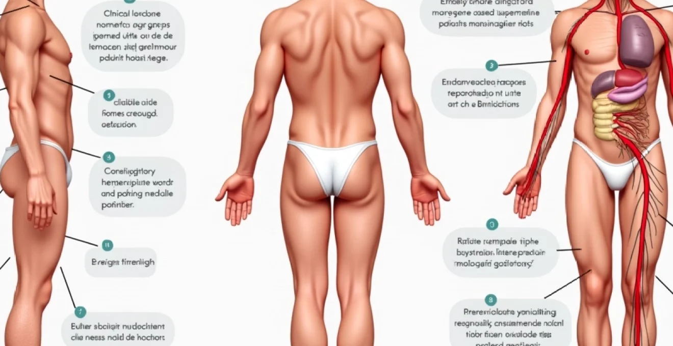
Anatomical structure and location of palpable lymph nodes
The human lymphatic system contains approximately 800 lymph nodes distributed throughout the body, though only those positioned superficially can be detected through physical examination. These bean-shaped structures cluster at strategic anatomical locations where lymphatic vessels converge, particularly around major blood vessel junctions. The most commonly palpable lymph node groups include the cervical chains, axillary clusters, inguinal regions, and supraclavicular areas.
Superficial cervical chain palpation techniques
The cervical lymph node chains represent the most frequently examined and easily palpable nodes in clinical practice. These structures are organised into anterior and posterior chains, with additional submental, submandibular, and preauricular groups forming a comprehensive network around the head and neck region. Proper palpation technique involves using the fingertips in gentle, circular motions whilst systematically examining each anatomical region in a consistent sequence.
The anterior cervical chain follows the path of the internal jugular vein, whilst the posterior chain aligns with the spinal accessory nerve and posterior border of the sternocleidomastoid muscle. During examination, slight head tilting towards the side being palpated helps relax the overlying muscle tissue, facilitating detection of enlarged nodes. Normal cervical lymph nodes may occasionally be palpable in thin individuals, particularly in the submandibular and upper cervical regions.
Axillary lymph node groups and anatomical boundaries
The axillary lymph node complex comprises five distinct groups: central, medial (pectoral), lateral (humeral), posterior (subscapular), and apical (infraclavicular) nodes. This arrangement creates a three-dimensional network that drains lymphatic fluid from the upper extremity, chest wall, and breast tissue. Effective examination requires proper positioning with slight arm elevation to relax the overlying musculature whilst maintaining access to the axillary space.
The central group, located in the apex of the axilla, represents the most commonly palpable nodes under normal circumstances. These structures can occasionally be felt in individuals with minimal subcutaneous tissue, particularly during periods of immune activation. The examination technique involves gentle pressure against the chest wall whilst moving systematically through each anatomical compartment of the axillary space.
Inguinal node distribution along saphenous vein pathways
Inguinal lymph nodes are arranged in superficial and deep groups, with the superficial nodes forming horizontal and vertical chains along the inguinal crease and great saphenous vein respectively. These nodes drain the lower extremity, external genitalia, and lower abdominal wall, making them particularly reactive to infections in these regions. The horizontal group lies parallel to the inguinal ligament, whilst the vertical group follows the medial aspect of the upper thigh.
Normal inguinal lymph nodes are more frequently palpable than nodes in other regions, particularly in active individuals who may experience minor trauma or infections affecting the lower extremities. The superficial location and relatively larger size of these nodes contribute to their detectability during routine self-examination or clinical assessment.
Supraclavicular fossa examination methods
The supraclavicular lymph nodes occupy the hollow areas above the clavicles, with particular clinical significance attached to the left supraclavicular fossa, known as Virchow’s node when enlarged. These nodes drain extensive regions including the thorax, abdomen, and left upper extremity through the thoracic duct. Examination requires specific positioning with shoulder elevation and forward flexion to create optimal access to these deeply situated structures.
Palpation technique involves placing fingers in the supraclavicular fossa whilst the patient hunches their shoulders forward, creating relaxation of the overlying skin and muscle. Normal supraclavicular nodes are rarely palpable, making any detectable enlargement in this region clinically significant and warranting further investigation.
Normal physiological parameters for lymph node detection
Understanding the normal characteristics of palpable lymph nodes requires knowledge of size thresholds, consistency patterns, mobility features, and temperature variations that distinguish physiological from pathological lymphadenopathy. These parameters provide clinicians and individuals with objective criteria for assessing lymph node significance and determining appropriate follow-up measures.
Size thresholds in millimetres for healthy nodes
Normal lymph nodes typically measure less than 10 millimetres in diameter, though this threshold varies by anatomical location and individual factors. Cervical and axillary nodes may reach 15 millimetres whilst remaining within normal limits, particularly in younger individuals with active immune systems. Inguinal nodes demonstrate the greatest size variation, with measurements up to 20 millimetres occasionally considered normal in healthy adults.
The assessment of lymph node size requires consistent measurement techniques and consideration of bilateral comparisons. Nodes that exceed established size thresholds or demonstrate progressive enlargement over time warrant clinical evaluation, regardless of associated symptoms. However, size alone cannot definitively distinguish benign from malignant lymphadenopathy , necessitating evaluation of additional characteristics.
Consistency and mobility characteristics in benign states
Normal and reactive lymph nodes typically exhibit a soft to moderately firm consistency with preserved mobility when palpated. These characteristics reflect the normal architecture of lymphoid tissue and the absence of infiltrative processes that might alter nodal structure. Benign nodes should move freely beneath the examining fingers and demonstrate distinct borders without fixation to surrounding tissues.
The consistency of normal lymph nodes can be compared to the firmness of the tip of the nose or the muscle tissue at the base of the thumb. Nodes that feel hard, rock-like, or demonstrate irregular surfaces may indicate pathological processes requiring further investigation. Similarly, fixation to underlying structures or skin involvement suggests advanced pathological changes.
Temperature variations during active immune response
Lymph nodes engaged in active immune responses may demonstrate increased warmth and tenderness compared to quiescent nodes. This inflammatory response reflects increased metabolic activity and vascular flow associated with immune cell proliferation and activation. The presence of warmth and tenderness often indicates benign reactive processes rather than malignant involvement.
Temperature assessment requires comparison with surrounding tissue and contralateral nodes when possible. Acute inflammatory conditions typically produce warm, tender lymphadenopathy that resolves as the underlying process subsides. Conversely, malignant lymphadenopathy often presents as painless, non-tender nodal enlargement without associated temperature changes.
Bilateral symmetry assessment in routine examination
Bilateral examination provides crucial comparative information for assessing lymph node significance. Normal anatomical variation can result in asymmetric lymph node palpability, but dramatic differences between sides warrant investigation. Systematic bilateral comparison helps distinguish constitutional variation from pathological processes affecting specific drainage regions.
The examination technique should include simultaneous bilateral palpation when anatomically feasible, allowing direct comparison of size, consistency, and mobility characteristics. Unilateral lymphadenopathy may reflect localised pathological processes, whilst bilateral involvement often suggests systemic conditions or widespread disease processes.
Clinical differentiation between normal and pathological lymphadenopathy
Distinguishing normal lymph node palpability from pathological lymphadenopathy requires systematic evaluation of multiple clinical parameters including onset pattern, associated symptoms, response to treatment, and epidemiological factors. This comprehensive assessment enables appropriate clinical decision-making and guides the need for further investigation or specialist referral.
Reactive hyperplasia following viral upper respiratory infections
Viral upper respiratory infections commonly trigger reactive lymphadenopathy in cervical node chains, representing normal immune system activation rather than pathological disease. This reactive hyperplasia typically develops within days of symptom onset and demonstrates characteristic patterns of tenderness, mobility, and gradual resolution following infection clearance.
The lymphadenopathy associated with viral infections usually affects multiple nodes within the drainage region of the infected tissue. Nodes remain mobile, moderately tender, and demonstrate soft to firm consistency throughout the reactive phase. Resolution typically occurs within 2-4 weeks following symptom resolution, though some residual palpability may persist for several additional weeks.
Bacterial infections can produce more dramatic lymphadenopathy with greater tenderness, warmth, and potential for suppuration. Streptococcal pharyngitis, dental abscesses, and skin infections commonly produce reactive cervical lymphadenopathy that responds appropriately to antimicrobial therapy. The temporal relationship between infection and lymphadenopathy provides important diagnostic clues.
Malignant transformation indicators using virchow’s node assessment
Virchow’s node, located in the left supraclavicular fossa, represents a sentinel site for detecting malignant disease affecting thoracic and abdominal organs. Enlargement of this node, termed Troisier’s sign, may indicate gastric, pancreatic, pulmonary, or other intra-abdominal malignancies with lymphatic spread. The anatomical connections through the thoracic duct make this location particularly significant for detecting occult malignancy.
Any palpable supraclavicular lymphadenopathy, particularly on the left side, requires prompt medical evaluation due to its association with serious underlying pathology.
Malignant lymphadenopathy typically demonstrates characteristic features including progressive enlargement, firm to hard consistency, fixation to surrounding structures, and absence of tenderness. These nodes may feel irregular, demonstrate poor mobility, and fail to respond to anti-inflammatory treatments or antibiotic therapy when infection is suspected.
Inflammatory conditions mimicking neoplastic lymph node changes
Various inflammatory conditions can produce lymphadenopathy that mimics malignant disease, creating diagnostic challenges requiring careful clinical assessment. Sarcoidosis, autoimmune disorders, and chronic inflammatory conditions may produce firm, enlarged lymph nodes that persist for extended periods without obvious infectious triggers.
Rheumatoid arthritis, systemic lupus erythematosus, and other connective tissue disorders commonly produce generalised lymphadenopathy as part of their systemic inflammatory response. These conditions typically affect multiple node groups simultaneously and demonstrate fluctuating courses corresponding to disease activity. Additional systemic symptoms and laboratory abnormalities usually provide diagnostic clues.
Cat scratch disease, toxoplasmosis, and other specific infections can produce persistent lymphadenopathy that may raise concern for malignancy. These conditions often affect specific anatomical regions corresponding to the route of infection and may require specialised diagnostic testing for definitive identification.
Systemic diseases manifesting through generalised lymphadenopathy
Generalised lymphadenopathy affecting multiple anatomical regions simultaneously often indicates systemic disease processes requiring comprehensive evaluation. Haematological malignancies, including lymphoma and leukaemia, commonly present with widespread lymph node involvement accompanied by constitutional symptoms such as fever, night sweats, and unexplained weight loss.
Infectious mononucleosis, HIV infection, and other viral syndromes can produce dramatic generalised lymphadenopathy in previously healthy individuals. These conditions typically affect younger demographics and may present with characteristic symptom complexes including pharyngitis, hepatosplenomegaly, and atypical lymphocytosis.
The presence of constitutional symptoms alongside lymphadenopathy significantly increases the likelihood of serious underlying pathology and warrants prompt medical evaluation. B symptoms, including unexplained fever, drenching night sweats, and weight loss exceeding 10% of body weight, are particularly concerning when accompanied by lymphadenopathy.
Age-related variations in lymph node palpability
Age significantly influences lymph node palpability, with children and young adults demonstrating greater baseline lymph node detectability compared to older individuals. This variation reflects differences in immune system activity, body composition, and exposure patterns to infectious agents throughout the lifespan. Understanding these age-related differences helps contextualise lymph node findings and guide appropriate clinical responses.
Children frequently have palpable lymph nodes in multiple anatomical regions as a normal finding related to ongoing immune system development and frequent exposure to novel antigens. Cervical, axillary, and inguinal lymphadenopathy measuring up to 15-20 millimetres may be entirely normal in healthy children, particularly those attending daycare or school settings with high infectious disease exposure rates.
Adolescents and young adults maintain relatively active immune systems that may produce readily palpable lymph nodes during minor infections or immune challenges. The lymphoid tissue reaches peak activity during this life stage, making reactive lymphadenopathy more common and pronounced compared to older adults. However, persistent or progressive lymphadenopathy in young adults requires careful evaluation due to the peak incidence of certain lymphomas in this demographic.
Older adults typically demonstrate decreased lymph node palpability due to age-related immune system changes, reduced lymphoid tissue mass, and altered body composition. When lymphadenopathy does occur in elderly individuals, it carries greater clinical significance and requires thorough investigation due to increased malignancy risk and decreased ability to mount robust immune responses to infections.
Pregnancy and hormonal changes can influence lymph node palpability through effects on immune system function and vascular dynamics. Breast tissue changes during pregnancy may make axillary lymph nodes more readily detectable, whilst hormonal influences on immune function may alter reactive patterns to infections or other stimuli.
Professional medical assessment protocols and red flag symptoms
Professional medical assessment of lymphadenopathy follows systematic protocols designed to identify high-risk features requiring urgent investigation whilst avoiding unnecessary anxiety and testing for benign conditions. These protocols incorporate epidemiological factors, clinical presentation patterns, and response to initial interventions to guide appropriate management decisions.
Red flag symptoms that mandate immediate medical evaluation include rapid lymph node enlargement over days to weeks, nodes exceeding 25-30 millimetres in diameter, fixation to surrounding structures, and constitutional symptoms suggesting systemic disease. The presence of multiple concerning features significantly increases the probability of serious underlying pathology requiring urgent investigation.
Lymph nodes that continue to enlarge despite appropriate antibiotic treatment for suspected infection require further evaluation to exclude malignant disease or unusual infectious processes.
The clinical assessment includes detailed history-taking focused on symptom onset, associated constitutional symptoms, recent infections, travel history, occupational exposures, and medication use. Physical examination extends beyond the palpable lymphadenopathy to include evaluation of all accessible node groups, assessment for hepatosplenomegaly, and examination of potential primary sites of infection or malignancy.
Laboratory investigations may include complete blood count with differential, inflammatory markers, and specific serological tests based on clinical suspicion. Imaging studies using ultrasound, CT, or MRI can provide additional characterisation of lymph node architecture and identify deep-seated adenopathy not detectable through physical examination. When clinical suspicion remains high despite negative initial investigations, tissue sampling through fine-needle aspiration or excisional biopsy may be necessary.
The concept of watchful waiting applies to lymphadenopathy with benign characteristics in appropriate clinical contexts. This approach involves systematic monitoring with defined follow-up intervals and clear criteria for escalating investigation if concerning changes develop. Patient education about warning signs and appropriate follow-up timing is essential for successful implementation of watchful waiting strategies.
Self-examination techniques and monitoring strategies for patient education
Effective self-examination of lymph nodes requires proper technique, systematic approach, and understanding of normal variations to avoid unnecessary anxiety whilst maintaining appropriate vigilance for concerning changes. Patient education should emphasise the importance of establishing baseline familiarity with normal lymph node patterns and recognising significant changes that warrant medical attention.
The recommended self-examination technique involves using the fingertips of the opposite hand to perform gentle, circular motions over lymph node regions. Excessive pressure should be avoided as it may obscure subtle findings or create discomfort that interferes with accurate assessment. The examination should be performed in consistent lighting with the individual in a relaxed position that allows optimal access to each anatom
ical region.
Monthly self-examination is generally recommended for individuals at higher risk of lymphoma or those with previous lymphadenopathy. However, excessive or compulsive checking can lead to anxiety and false alarms from normal anatomical variations or temporary reactive changes. The examination should be performed at the same time each month, ideally after bathing when the skin is clean and supple.
Documentation of findings through simple notes or photographs can help track changes over time and provide valuable information for healthcare providers. Recording the size, location, consistency, and any associated symptoms creates a baseline reference for detecting significant alterations. This systematic approach helps distinguish normal fluctuations from progressive changes requiring medical attention.
Patient education must emphasise the difference between normal lymph node palpability and concerning lymphadenopathy. Understanding that small, mobile, soft lymph nodes are often normal helps prevent unnecessary anxiety whilst maintaining appropriate vigilance for worrisome changes. Clear criteria for seeking medical attention should be established, including progressive enlargement, development of constitutional symptoms, or persistence beyond expected timeframes for reactive processes.
The self-examination should include visual inspection for obvious asymmetry or skin changes overlying lymph node regions. Lymph nodes affected by infection may produce visible swelling, whilst malignant involvement occasionally causes skin changes including erythema, peau d’orange appearance, or ulceration. These visual changes often precede significant palpable alterations and warrant immediate medical evaluation.
Timing of self-examination relative to illness or vaccination is important for accurate interpretation of findings. Recent infections, immunisations, or dental procedures may cause temporary lymphadenopathy that resolves spontaneously. Understanding these temporal relationships helps distinguish expected reactive changes from pathological processes requiring intervention.
Effective patient education includes practical demonstrations of proper palpation technique during clinical encounters. Healthcare providers should guide patients through systematic examination of their own lymph nodes, allowing them to experience the normal feel and location of these structures. This hands-on approach builds confidence and competence for accurate self-assessment whilst addressing individual anatomical variations that might cause confusion.
The psychological aspects of lymph node self-examination require careful consideration, as excessive worry about lymphadenopathy can significantly impact quality of life. Balanced education should acknowledge legitimate concerns whilst providing reassurance about normal findings and clear guidance about when professional evaluation is warranted. Empowering patients with knowledge and practical skills reduces anxiety whilst maintaining appropriate health vigilance.
Technology integration through smartphone applications or online tracking tools can enhance self-examination effectiveness for motivated individuals. These tools can provide reminders for regular examinations, standardised documentation formats, and educational resources about normal versus abnormal findings. However, technology should supplement rather than replace proper clinical evaluation when concerning features are identified.
Special populations including immunocompromised individuals, cancer survivors, and those with chronic inflammatory conditions require modified self-examination strategies tailored to their specific risk profiles. These individuals may need more frequent monitoring, lower thresholds for seeking medical attention, and enhanced coordination with their healthcare teams to distinguish disease-related changes from treatment effects or disease progression.