The moment your healthcare provider removes that bulky walking boot, you might expect immediate relief and a swift return to normal activities. However, many patients experience surprising discomfort and pain following boot removal, leading to concerns about incomplete healing or complications. This phenomenon is more common than you might think, affecting approximately 70% of patients who have undergone prolonged immobilisation with controlled ankle motion (CAM) boots. Understanding the underlying mechanisms behind post-immobilisation pain can help you navigate the recovery process with realistic expectations and appropriate management strategies. The transition from protected weight-bearing to normal ambulation represents a critical phase in lower extremity injury rehabilitation, requiring careful attention to both physical and psychological factors that influence pain perception.
Understanding Post-Immobilisation pain syndrome after walking boot removal
Post-immobilisation pain syndrome encompasses a complex array of symptoms that emerge following the removal of external support devices like walking boots. This condition affects multiple anatomical structures simultaneously, creating a cascade of physiological changes that can persist for weeks or months after boot discontinuation. The syndrome represents your body’s natural response to prolonged disuse and the subsequent reactivation of previously protected tissues.
Research indicates that approximately 30% of patients continue to experience significant discomfort three months after walking boot removal, with symptoms ranging from mild stiffness to severe activity-limiting pain. The duration and severity of these symptoms correlate strongly with the initial injury type, immobilisation period, and individual patient factors such as age, activity level, and compliance with rehabilitation protocols. Understanding this timeline helps set realistic expectations for your recovery journey.
Pathophysiology of muscle atrophy and joint stiffness following controlled ankle motion (CAM) boot treatment
The physiological changes that occur during walking boot immobilisation create a perfect storm for post-removal pain. Muscle atrophy begins within 72 hours of immobilisation, with calf muscles losing approximately 20-30% of their strength after just six weeks of disuse. The gastrocnemius and soleus muscles, in particular, undergo significant changes in fibre composition and cross-sectional area, leading to weakness and altered biomechanics upon return to activity.
Joint stiffness develops through multiple mechanisms, including capsular contracture, ligament shortening, and the formation of intra-articular adhesions. The ankle joint, designed for frequent movement, becomes particularly susceptible to these changes when held in a fixed position for extended periods. Synovial fluid production decreases, cartilage nutrition suffers, and the joint capsule thickens, creating mechanical restrictions that contribute to pain and movement limitations.
Distinguishing normal rehabilitation discomfort from pathological pain patterns
Normal post-boot removal discomfort typically manifests as mild to moderate aching, stiffness that improves with gentle movement, and fatigue during initial weight-bearing activities. This type of pain usually follows a predictable pattern, decreasing gradually over several weeks as tissues adapt to increased demands. You might experience sharp, brief sensations when moving your ankle into previously restricted positions, but these should not be severe or persistent.
Pathological pain patterns, conversely, present as severe, constant discomfort that interferes with sleep and daily activities. This pain often includes burning sensations, extreme sensitivity to light touch, or disproportionate responses to minimal stimuli. If you experience swelling that doesn’t respond to elevation, skin colour changes, or pain that worsens rather than improves over the first few weeks post-boot removal, these may indicate complications requiring immediate medical attention.
The key distinction lies in the pain’s response to gentle movement and time – normal rehabilitation discomfort gradually improves with appropriate activity, while pathological pain remains constant or worsens despite conservative management.
Timeline expectations for Post-Boot removal recovery in lower extremity injuries
Recovery timelines vary significantly based on injury type, immobilisation duration, and individual factors. For simple ankle sprains requiring 4-6 weeks of boot wear, you can expect initial discomfort to resolve within 2-3 weeks of boot removal, with full functional recovery achieved within 6-8 weeks. More complex injuries, such as ankle fractures requiring 8-12 weeks of immobilisation, may require 3-6 months for complete rehabilitation.
The first week post-boot removal typically involves the most significant discomfort as your tissues begin adapting to increased movement and weight-bearing demands. Stiffness is usually most pronounced in the morning and improves throughout the day with gentle activity. By the second week, you should notice gradual improvements in range of motion and a reduction in baseline pain levels, though activity-related discomfort may persist.
Impact of immobilisation duration on soft tissue adaptation and pain response
The duration of immobilisation directly correlates with the severity and persistence of post-removal symptoms. Studies demonstrate that immobilisation periods exceeding eight weeks result in significantly greater muscle atrophy, joint stiffness, and bone density loss compared to shorter durations. Each additional week of immobilisation can extend the recovery timeline by approximately 1.5-2 weeks, highlighting the importance of minimising immobilisation time when medically appropriate.
Connective tissue adaptation occurs throughout the immobilisation period, with tendons and ligaments losing elasticity and strength. The Achilles tendon, in particular, undergoes significant shortening during walking boot wear, contributing to posterior heel pain and restricted dorsiflexion upon boot removal. These adaptations require targeted interventions to restore normal tissue properties and function.
Clinical assessment of Post-Walking boot pain manifestations
Comprehensive assessment of post-walking boot pain requires systematic evaluation of multiple anatomical structures and functional domains. Healthcare providers must consider the complex interplay between mechanical factors, neurological sensitisation, and psychological influences that contribute to the overall pain experience. This assessment forms the foundation for developing targeted treatment strategies that address the specific manifestations present in each individual case.
The clinical presentation of post-boot pain varies considerably among patients, with some experiencing focal symptoms while others develop generalised discomfort throughout the lower extremity. Understanding these diverse manifestations enables more precise diagnosis and treatment planning, ultimately leading to better outcomes and reduced recovery times.
Identifying achilles tendon tightness and posterior heel pain after boot discontinuation
Achilles tendon complications represent one of the most common sources of post-boot pain, affecting up to 85% of patients following prolonged immobilisation. The tendon undergoes adaptive shortening during boot wear, particularly when heel elevation wedges are used or when the foot is positioned in plantarflexion. This shortening creates significant tension when attempting to return to normal dorsiflexion ranges, resulting in posterior heel pain and functional limitations.
Clinical assessment reveals restricted ankle dorsiflexion, often limited to 5-10 degrees compared to the unaffected limb’s normal 15-20 degrees. Patients frequently report sharp, pulling sensations along the posterior calf and heel during walking, particularly when ascending stairs or walking uphill. The pain typically worsens with initial weight-bearing after periods of rest, a phenomenon known as post-static dyskinesia.
Evaluating plantar fascia contracture and arch discomfort following immobilisation
Plantar fascia contracture develops insidiously during walking boot immobilisation, as the tissue adapts to the altered foot position and reduced loading patterns. The fascia loses its normal elasticity and becomes shortened, leading to increased tension across the arch during weight-bearing activities. This contracture contributes significantly to morning stiffness and the characteristic “first-step pain” experienced by many patients following boot removal.
Arch discomfort manifests as a deep, aching sensation that typically worsens with prolonged standing or walking on hard surfaces. The pain often radiates from the heel towards the midfoot and may be accompanied by a sensation of tightness or pulling along the bottom of the foot. Assessment techniques include palpation of the plantar fascia insertion, evaluation of passive dorsiflexion with knee extension, and functional testing of arch support during weight-bearing activities.
Recognising ankle joint capsule restriction and associated morning stiffness
Joint capsule restriction represents a significant contributor to post-boot pain and functional limitation. The ankle joint capsule thickens and contracts during immobilisation, creating mechanical blocks to normal range of motion in multiple planes. This restriction affects both talocrural and subtalar joint mobility, leading to compensatory movement patterns that can perpetuate pain and dysfunction.
Morning stiffness serves as a hallmark symptom of capsular restriction, with patients reporting severe difficulty initiating movement upon awakening. The stiffness typically requires 15-30 minutes of gentle movement to improve, though complete resolution may take several hours. Joint capsule restriction can be assessed through passive range of motion testing, joint glide evaluation, and functional movement screens that highlight movement compensations.
Assessing calf muscle weakness and Exercise-Induced soreness patterns
Calf muscle weakness following walking boot removal creates a cascade of functional deficits that contribute to ongoing pain and movement dysfunction. The gastrocnemius and soleus muscles lose both strength and endurance capacity during immobilisation, with weakness persisting for months after boot discontinuation. This weakness affects push-off power during walking, contributes to altered gait mechanics, and increases the risk of re-injury.
Exercise-induced soreness patterns help differentiate between normal muscle reconditioning and pathological pain responses. Normal reconditioning soreness appears 24-48 hours after activity, responds well to gentle movement and heat application, and gradually decreases in intensity over several days. Pathological responses include immediate severe pain during activity, persistent soreness lasting more than 72 hours, or pain that worsens with continued activity despite proper progression.
Evidence-based rehabilitation protocols for Post-Boot pain management
Effective management of post-walking boot pain requires a systematic, evidence-based approach that addresses the multiple contributing factors simultaneously. Modern rehabilitation protocols emphasise early mobilisation, graded exposure to loading, and neuromuscular re-education to optimise tissue healing while minimising pain and dysfunction. These protocols must be individualised based on the specific injury type, patient characteristics, and presenting symptoms to achieve optimal outcomes.
The rehabilitation process typically progresses through distinct phases, each with specific goals and interventions designed to address the evolving needs of healing tissues. Research demonstrates that structured, progressive rehabilitation programmes reduce pain intensity by an average of 60-70% within the first month post-boot removal, while also improving functional outcomes and reducing the risk of long-term complications.
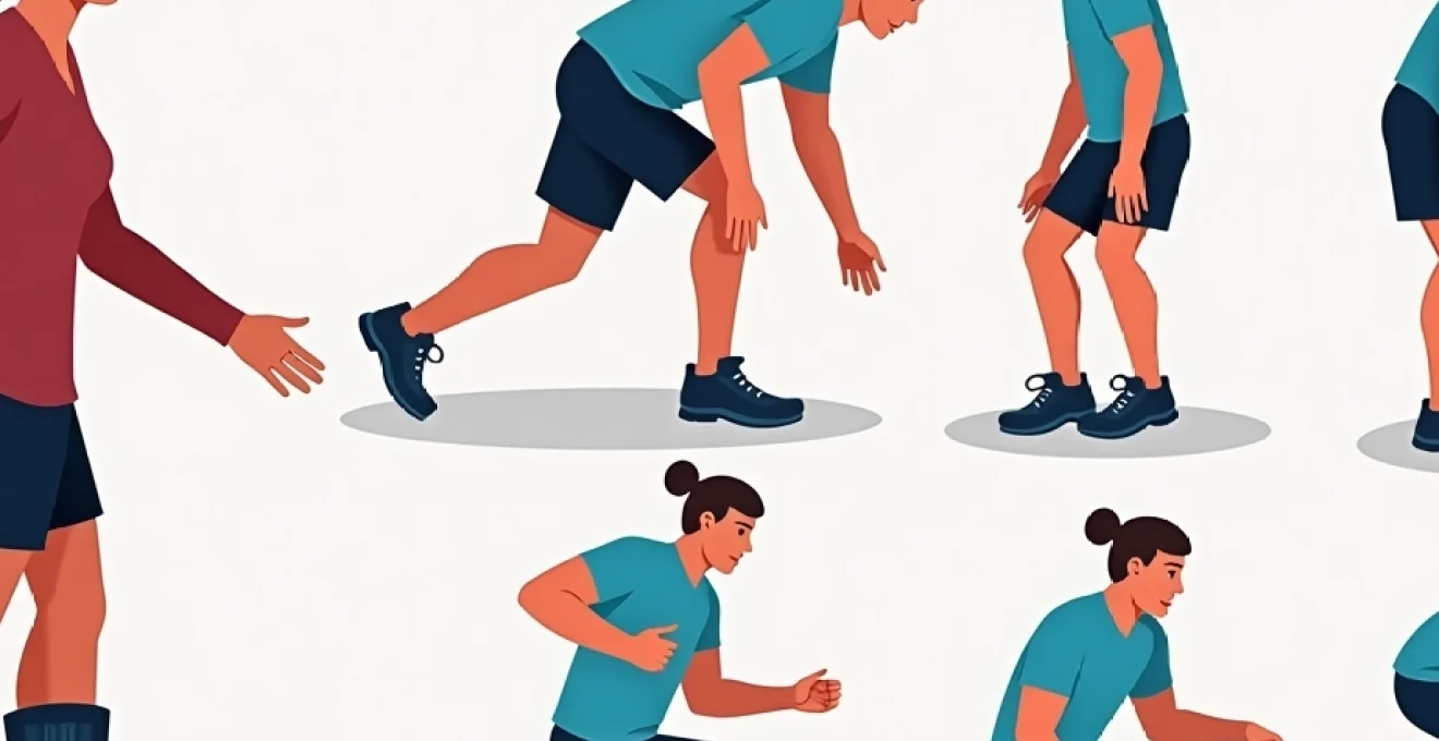
Progressive Weight-Bearing protocols using Heel-to-Toe gait retraining
Progressive weight-bearing protocols form the cornerstone of post-boot rehabilitation, providing graduated exposure to loading forces while protecting healing tissues from excessive stress. These protocols typically begin with partial weight-bearing at 25-50% of body weight, progressing incrementally based on pain response and tissue tolerance. Heel-to-toe gait retraining helps restore normal walking mechanics that may have been disrupted during the period of altered mobility.
Gait retraining focuses on re-establishing proper heel strike, midstance stability, and push-off mechanics through the affected limb. Patients learn to distribute weight evenly across the foot, maintain proper ankle alignment, and coordinate the complex sequence of muscle activations required for normal walking. Visual feedback techniques, such as mirror training or video analysis, help patients recognise and correct movement compensations that may perpetuate pain and dysfunction.
Targeted stretching regimens for gastrocnemius and soleus complex restoration
Targeted stretching regimens address the specific soft tissue restrictions that develop during walking boot immobilisation. The gastrocnemius and soleus muscles require different stretching approaches due to their distinct anatomical characteristics and functional roles. Gastrocnemius stretching is performed with the knee extended to target this bi-articular muscle effectively, while soleus stretching requires knee flexion to isolate this deeper muscle group.
Stretching protocols typically involve multiple daily sessions lasting 30-60 seconds per repetition, with 3-5 repetitions per muscle group. The intensity should be sufficient to create mild tension without causing sharp pain or protective muscle guarding. Progressive stretching techniques, including contract-relax methods and dynamic stretching, can enhance effectiveness while reducing the risk of tissue irritation during the sensitive post-boot period.
Consistency in stretching frequency proves more important than intensity – gentle, frequent stretching sessions throughout the day yield superior results compared to aggressive, infrequent sessions that may cause tissue irritation.
Proprioceptive training exercises to address balance deficits and ankle instability
Proprioceptive deficits develop rapidly during periods of immobilisation, with balance control and joint position sense showing significant deterioration within just a few weeks of walking boot use. These deficits contribute to increased fall risk, functional instability, and the development of compensatory movement patterns that can perpetuate pain and dysfunction. Proprioceptive training exercises help restore these essential sensorimotor functions through progressive challenges to the balance and stability systems.
Training progressions typically begin with static balance activities on stable surfaces, advancing to dynamic balance challenges on unstable surfaces such as foam pads or balance boards. Single-leg stance exercises, weight-shifting activities, and perturbation training help restore confidence in the affected limb while improving neuromuscular control. The incorporation of cognitive challenges, such as dual-task activities, enhances the functional relevance of proprioceptive training.
Gradual return to activities of daily living through functional movement patterns
Functional movement pattern training bridges the gap between basic rehabilitation exercises and real-world activity demands. This approach emphasises movements that replicate common daily activities such as stair climbing, squatting, and walking on uneven surfaces. By practising these functional patterns in a controlled environment, patients develop the confidence and competence needed for safe return to normal activities.
The progression of functional activities follows a systematic hierarchy, beginning with basic movements in stable environments and advancing to complex, multi-planar activities in challenging conditions. Task-specific training addresses the unique demands of each patient’s lifestyle and occupational requirements, ensuring that rehabilitation outcomes translate effectively to real-world function. Regular assessment of functional progress helps guide advancement decisions and identifies areas requiring additional attention.
Red flag symptoms requiring immediate medical evaluation
While post-walking boot pain is often a normal part of the recovery process, certain symptoms indicate serious complications that require immediate medical attention. Recognising these red flag symptoms can prevent potentially devastating consequences and ensure appropriate escalation of care when necessary. Healthcare providers emphasise the importance of patient education regarding these warning signs, as early recognition and treatment significantly improve outcomes for serious complications.
The distinction between normal rehabilitation discomfort and pathological symptoms can be challenging for patients to navigate independently. Red flag symptoms typically present with characteristic patterns that differ markedly from expected recovery trajectories. These symptoms may indicate complications such as complex regional pain syndrome, deep vein thrombosis, compartment syndrome, or infection, all of which require prompt medical intervention to prevent long-term sequelae.
Severe, disproportionate pain that seems excessive relative to the original injury or expected healing timeline represents one of the most significant red flags. This pain often described as burning, electric, or crushing in nature, may be accompanied by extreme sensitivity to light touch or temperature changes. The pain typically fails to respond to standard pain management strategies and may actually worsen with gentle movement or rehabilitation activities.
Circulatory compromise manifests through several concerning symptoms that require immediate evaluation. These include persistent swelling that fails to respond to elevation, skin colour changes ranging from pallor to cyanosis, decreased skin temperature compared to the unaffected limb, and absent or diminished pulses. Deep vein thrombosis presents particular risks following prolonged immobilisation, with symptoms including unilateral calf pain, warmth, swelling, and visible venous distension.
Neurological symptoms indicating potential nerve compression or injury include persistent numbness or tingling that doesn’t improve with position changes, progressive weakness in muscles not directly affected by the original injury, or the development of foot drop or other focal neurological deficits. These symptoms may indicate compartment syndrome, nerve entrapment, or other serious complications requiring urgent intervention.
Infection-related red flags encompass local signs such as increasing redness, warmth, purulent drainage, or foul odour around previous surgical sites or areas of skin breakdown. Systemic symptoms including fever, chills, malaise, or unexplained fatigue may indicate spreading infection requiring immediate antibiotic treatment. The risk of infection increases in patients with diabetes, immunocompromise, or poor circulation.
Long-term prognosis and prevention of chronic Post-Immobilisation complications
The long-term prognosis for patients experiencing post-walking boot pain is generally favourable, with the majority achieving substantial improvement within three to six months of boot removal. Research indicates that 85-90% of patients return to their pre
-injury activity level within twelve months of completing appropriate rehabilitation. However, the recovery trajectory varies significantly based on adherence to rehabilitation protocols, underlying health conditions, and the development of compensatory movement patterns during the recovery phase.
Chronic post-immobilisation complications affect approximately 15-25% of patients, with the most common long-term issues including persistent joint stiffness, recurrent pain episodes, and functional limitations that interfere with desired activities. These complications often stem from inadequate initial rehabilitation, premature return to high-impact activities, or the development of fear-avoidance behaviours that perpetuate movement restrictions and muscle weakness.
Prevention strategies focus on early identification of risk factors and proactive intervention to address emerging problems before they become entrenched. Patients with longer immobilisation periods, multiple previous injuries, or pre-existing conditions such as diabetes or arthritis face higher risks for chronic complications. Regular monitoring during the first six months post-boot removal allows healthcare providers to identify subtle changes in symptoms or function that may indicate the need for intervention adjustments.
Activity modification plays a crucial role in preventing long-term complications while supporting continued healing. Gradual progression of loading and impact activities allows tissues to adapt appropriately without overwhelming their current capacity. Patients who attempt to return to pre-injury activity levels too quickly often experience setbacks that can prolong recovery and increase the risk of chronic pain development.
The key to preventing chronic complications lies in balancing appropriate challenge with adequate recovery time – pushing too hard too fast often leads to setbacks that extend the overall recovery timeline.
Patient education strategies for managing expectations during walking boot weaning process
Effective patient education forms the foundation of successful walking boot weaning, significantly influencing both short-term recovery outcomes and long-term functional results. Research demonstrates that patients who receive comprehensive education about the weaning process experience 40% less anxiety and demonstrate better adherence to rehabilitation protocols compared to those receiving minimal instruction. The education process must address both the physiological aspects of recovery and the psychological challenges associated with transitioning from protected mobility to independent function.
The weaning process typically spans 2-4 weeks, during which patients gradually reduce their reliance on the walking boot while increasing normal weight-bearing activities. This transition period represents a critical window where appropriate expectations and understanding of normal symptoms can prevent unnecessary anxiety and promote optimal recovery behaviours. Education should begin before boot removal to prepare patients mentally and physically for the challenges ahead.
Expectation management requires honest discussion about the reality of post-boot symptoms and recovery timelines. Many patients expect immediate return to normal function upon boot removal, leading to disappointment and concern when they experience ongoing discomfort. Healthcare providers should clearly explain that some degree of pain, stiffness, and functional limitation is normal and expected during the initial weeks following boot discontinuation.
Pain education helps patients understand the difference between harmful pain that requires immediate attention and normal rehabilitation discomfort that indicates healing progress. Teaching patients to use numerical pain scales effectively allows for better communication with healthcare providers and more informed decision-making about activity progression. Patients should understand that mild to moderate pain that improves with gentle movement and decreases over time represents normal tissue adaptation, while severe, constant, or worsening pain may indicate complications.
Activity pacing strategies prevent the boom-bust cycle that often prolongs recovery and increases frustration. Patients learn to break activities into manageable segments, alternate between challenging and easier tasks, and recognise early signs of overexertion before symptoms become overwhelming. Pacing education includes specific guidance on walking distances, standing tolerance, and appropriate rest intervals based on individual capacity and healing status.
Home exercise program education ensures that patients can safely and effectively perform prescribed interventions between healthcare visits. This education includes proper technique demonstration, safety precautions, progression criteria, and troubleshooting common problems. Patients should understand how to modify exercises based on daily symptoms while maintaining overall progression toward recovery goals.
Return-to-activity guidelines provide clear criteria for resuming various daily, recreational, and occupational activities. These guidelines should be specific to individual patient goals and realistic about timelines for different activity levels. For example, return to walking for exercise may be appropriate within 2-3 weeks of boot removal, while return to running or jumping activities may require 2-3 months of progressive rehabilitation.
Warning sign recognition empowers patients to identify symptoms requiring immediate medical attention while reducing anxiety about normal recovery sensations. Education should cover red flag symptoms such as severe swelling, skin colour changes, numbness, or disproportionate pain, along with clear instructions for accessing healthcare when concerns arise. This knowledge helps patients feel confident in managing their recovery while ensuring appropriate medical oversight.
Support system utilisation acknowledges that recovery extends beyond the individual patient to include family members, employers, and other support networks. Education should address how family members can assist with home exercises, activity modifications, and emotional support during challenging phases of recovery. Workplace accommodations may be necessary during the transition period, requiring clear communication about functional limitations and expected improvement timelines.
Long-term maintenance strategies help patients understand that recovery extends beyond the formal rehabilitation period. Ongoing attention to flexibility, strength, and movement quality helps prevent re-injury and maintains optimal function. Patients should understand the importance of continuing key exercises even after formal therapy ends, along with strategies for maintaining activity levels that support continued ankle health.