Experiencing pressure behind the head and ears can be a perplexing and uncomfortable sensation that affects millions of people worldwide. This complex symptom often results from intricate interactions between anatomical structures, vascular systems, and neurological pathways that govern cranial pressure regulation. Understanding the underlying mechanisms behind these sensations requires examining the sophisticated network of bones, nerves, blood vessels, and soft tissues that work together to maintain proper head and ear function. The sensation of pressure in these regions can manifest as dull aching, throbbing, or intense discomfort that may significantly impact daily activities and quality of life.
Anatomical structures contributing to occipital and auricular pressure sensations
The complex anatomy of the posterior cranium and auricular region involves multiple interconnected structures that can contribute to pressure sensations. The occipital bone forms the back of the skull and houses critical neural pathways, while the temporal bones contain intricate ear structures that regulate both hearing and balance. These anatomical components work in harmony to process sensory information and maintain cranial stability, but when dysfunction occurs, they can generate significant discomfort.
Temporal bone architecture and mastoid process involvement
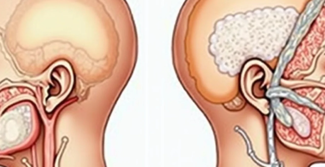
The temporal bone’s mastoid process represents a crucial anatomical landmark that frequently contributes to retroauricular pressure sensations. This honeycomb-like structure contains air cells that communicate directly with the middle ear space through the mastoid antrum. When inflammation or infection develops within these air cells, a condition known as mastoiditis can occur, creating intense pressure and pain behind the ear. The proximity of the mastoid process to important neural structures, including the facial nerve and various cranial nerve branches, means that pathology in this region can produce complex symptom patterns. Chronic inflammation within the mastoid air cells can also lead to bone erosion and complications affecting nearby structures, including the inner ear and intracranial compartments.
Trigeminal nerve pathway distribution in cranial pressure perception
The trigeminal nerve, specifically its auriculotemporal branch, plays a significant role in transmitting sensory information from the temporal region and external ear. This nerve pathway can become sensitised or compressed, leading to referred pain patterns that manifest as pressure sensations behind the ears and extending toward the occipital region. The auriculotemporal nerve’s close relationship with the temporomandibular joint means that TMJ dysfunction can directly impact this nerve’s function, creating a cascade of symptoms that include ear pressure, jaw pain, and temporal headaches. Understanding this neural pathway helps explain why seemingly unrelated conditions affecting the jaw can produce ear and head pressure symptoms.
Eustachian tube dysfunction and barometric pressure regulation
The Eustachian tubes serve as critical pressure-equalising mechanisms between the middle ear and nasopharynx. When these tubes fail to function properly, a condition known as Eustachian tube dysfunction develops, creating persistent pressure sensations behind the ears. This dysfunction can result from allergies, upper respiratory infections, or anatomical variations that prevent proper tube opening and closing. The resulting pressure imbalance can create a sensation of fullness, muffled hearing, and discomfort that extends beyond the ear itself. Chronic Eustachian tube dysfunction can lead to secondary complications including middle ear infections and tympanic membrane retraction, further exacerbating pressure symptoms.
Cervical vertebrae C1-C3 nerve root compression mechanisms
The upper cervical spine, particularly the C1, C2, and C3 vertebrae, houses nerve roots that contribute significantly to occipital and auricular sensation. The greater and lesser occipital nerves, which arise from the C2 and C3 nerve roots respectively, provide sensory innervation to the posterior scalp and can refer pain to the ear region when compressed or irritated. Cervical spine dysfunction, whether from poor posture, degenerative changes, or trauma, can create nerve root compression that manifests as pressure sensations extending from the neck to the back of the head and ears. This phenomenon, often termed cervicogenic headache , demonstrates the interconnected nature of spinal and cranial pain pathways.
Vascular pathophysiology behind cranial pressure manifestations
The vascular system supplying the posterior cranium and temporal regions represents a complex network of arteries and veins that must maintain precise pressure relationships to ensure proper brain function. Disruptions in this delicate balance can manifest as pressure sensations, headaches, and other uncomfortable symptoms. The cerebrovascular system’s intricate autoregulation mechanisms normally compensate for pressure variations, but when these systems become overwhelmed or dysfunctional, patients may experience significant discomfort.
Posterior cerebral artery hypertension and occipital lobe perfusion
Elevated pressure within the posterior cerebral artery circulation can create pulsatile pressure sensations in the occipital region. This condition often develops secondary to systemic hypertension but can also result from localised vascular pathology such as arterial stenosis or vascular malformations. The posterior cerebral arteries supply critical brain regions including the occipital lobes, and when perfusion pressure becomes elevated, patients may experience throbbing headaches, visual disturbances, and pressure sensations extending from the back of the head toward the ears. Vascular imaging studies frequently reveal abnormal flow patterns in these vessels when patients present with chronic occipital pressure symptoms.
Vertebral artery insufficiency syndrome in Neck-Head transition
The vertebral arteries traverse through the cervical vertebrae before joining to form the basilar artery, and compression or stenosis of these vessels can produce complex symptom patterns including occipital pressure. Vertebrobasilar insufficiency often manifests with posterior headaches, dizziness, and pressure sensations that worsen with certain neck positions or movements. This condition becomes increasingly common with advancing age as atherosclerotic changes narrow the vertebral arteries. The relationship between neck position and symptom severity often provides important diagnostic clues, as rotational or extension movements can further compromise already compromised blood flow.
Internal jugular vein compression and venous return obstruction
Impaired venous drainage from the cranial cavity can create increased intracranial pressure that manifests as diffuse head pressure, including sensations behind the ears and in the occipital region. Internal jugular vein compression can occur due to muscular tension, anatomical variations, or external compression from cervical spine pathology. When venous return becomes compromised, the resulting pressure buildup can create symptoms similar to those seen in idiopathic intracranial hypertension. Patients often report that their symptoms worsen when lying flat or with Valsalva manoeuvres, reflecting the impact of compromised venous drainage on intracranial pressure dynamics.
Carotid artery stenosis impact on temporal region blood flow
Stenosis of the carotid arteries, particularly the external carotid artery branches that supply the temporal region, can create altered perfusion patterns that contribute to temporal and retroauricular pressure sensations. The superficial temporal artery, a terminal branch of the external carotid, provides significant blood supply to the temporal region and scalp. When stenotic lesions develop proximally, compensatory vascular mechanisms may create altered flow patterns that patients perceive as pressure or throbbing sensations. Carotid ultrasound studies can reveal these vascular abnormalities and help guide appropriate treatment strategies.
Otolaryngological conditions causing retroauricular pressure
The ear, nose, and throat system represents an interconnected network where dysfunction in one area can create symptoms throughout the region. Retroauricular pressure often originates from specific otolaryngological pathology that disrupts normal pressure relationships or inflammatory processes within the temporal bone and surrounding structures.
Chronic otitis media with effusion and tympanic membrane tension
Chronic otitis media with effusion represents a persistent inflammatory condition where fluid accumulates behind the tympanic membrane without active infection. This fluid buildup creates constant pressure against the eardrum and surrounding structures, leading to sensations of fullness and pressure that can extend beyond the ear itself. The trapped fluid prevents normal tympanic membrane mobility and can create negative pressure within the middle ear space. Over time, this condition can lead to tympanic membrane retraction and secondary complications including hearing loss and chronic discomfort. Tympanometry testing typically reveals characteristic patterns that confirm the diagnosis and guide treatment decisions.
Temporomandibular joint disorder and referred otalgia
The temporomandibular joint’s close anatomical relationship to the ear means that TMJ dysfunction frequently produces referred pain and pressure sensations in the retroauricular region. The joint capsule shares innervation with structures surrounding the ear through the auriculotemporal nerve, creating a pathway for referred symptoms. Patients with TMJ disorders often experience ear pressure, particularly during jaw movements such as chewing or yawning. The masseter and temporalis muscles, when in spasm or chronically contracted, can create tension patterns that extend toward the ear and posterior head regions. Understanding this relationship helps explain why comprehensive TMJ treatment often resolves associated ear pressure symptoms.
Acoustic neuroma growth patterns and cranial nerve VIII compression
Acoustic neuromas, though rare, represent important causes of unilateral ear pressure and associated symptoms. These benign tumours grow along the eighth cranial nerve and can create pressure effects on surrounding structures within the cerebellopontine angle. As the tumour enlarges, it can compress not only the auditory and vestibular nerves but also affect nearby cranial nerves and brainstem structures. Early symptoms often include unilateral hearing loss, tinnitus, and a sensation of ear fullness or pressure. The slow growth pattern of these tumours means symptoms often develop gradually, making early diagnosis challenging. Magnetic resonance imaging remains the gold standard for acoustic neuroma detection and monitoring.
Ménière’s disease endolymphatic hydrops and pressure fluctuations
Ménière’s disease involves abnormal accumulation of endolymph within the inner ear, creating fluctuating pressure that produces characteristic symptoms including ear fullness, hearing loss, tinnitus, and vertigo. The endolymphatic hydrops creates mechanical pressure on delicate inner ear structures, disrupting both hearing and balance function. Patients often describe a sensation of ear fullness or pressure that may precede acute vertiginous episodes. The fluctuating nature of symptoms reflects the dynamic changes in endolymphatic pressure that characterise this condition. Dietary modifications, diuretic therapy, and other interventions aim to reduce endolymphatic pressure and stabilise inner ear function.
Neurological disorders manifesting as Occipital-Auricular pressure
Several neurological conditions can present with pressure sensations in the occipital and auricular regions, often reflecting dysfunction in pain processing pathways or direct neural compression. Occipital neuralgia represents one of the most common neurological causes of posterior head pressure, involving irritation or inflammation of the greater and lesser occipital nerves. These nerves emerge from the upper cervical spine and travel through the posterior scalp, and when compressed or irritated, they can produce sharp, shooting, or burning pain that may be accompanied by constant pressure sensations. The pain typically follows the nerve distribution pattern, extending from the suboccipital region toward the vertex of the scalp, and may include referred sensations to the ear region.
Trigeminal neuralgia, while typically affecting the facial region, can occasionally present with atypical patterns that include retroauricular pressure and discomfort. The trigeminal nerve’s complex distribution and connections with other cranial nerves can create referred pain patterns that extend beyond the traditional facial nerve territory. Migraine headaches frequently involve the temporal and occipital regions, creating pressure sensations that patients describe as band-like or vice-like compression around the head. The neurogenic inflammation associated with migraine can affect multiple cranial structures simultaneously, explaining the complex symptom patterns that often include ear pressure and occipital discomfort.
Cervicogenic headaches represent another important neurological cause of occipital pressure, arising from dysfunction in the upper cervical spine and its associated neural structures. The trigeminocervical complex creates anatomical connections between cervical afferents and trigeminal pathways, allowing cervical pathology to manifest as head and ear pressure. Post-traumatic headaches following concussion or whiplash injuries can also produce chronic pressure sensations, reflecting ongoing dysfunction in pain processing mechanisms and structural damage to cervical and cranial tissues.
Systemic medical conditions affecting cranial pressure homeostasis
Various systemic medical conditions can disrupt normal cranial pressure regulation, leading to persistent pressure sensations behind the head and ears. Hypertension represents one of the most significant systemic factors, as elevated blood pressure can directly affect cerebral perfusion and intracranial pressure dynamics. Chronic hypertension can lead to vascular remodelling that alters normal autoregulation mechanisms, creating pressure sensations and headaches that often localise to the occipital region. The relationship between blood pressure and head pressure symptoms often becomes apparent during periods of poor blood pressure control or medication adjustments.
Thyroid disorders, particularly hypothyroidism, can contribute to cranial pressure symptoms through multiple mechanisms including fluid retention, altered vascular function, and metabolic effects on neural tissue. Patients with thyroid dysfunction often report headaches and pressure sensations that improve with appropriate hormone replacement therapy. Sleep apnoea represents another systemic condition that can create cranial pressure symptoms through intermittent hypoxia and altered intracranial pressure patterns during sleep. The repetitive episodes of hypoxia and reoxygenation can lead to vascular dysfunction and chronic inflammation that manifests as morning headaches and pressure sensations.
Autoimmune conditions such as lupus, rheumatoid arthritis, and temporal arteritis can create cranial pressure symptoms through inflammatory mechanisms that affect vascular and neural structures. Temporal arteritis specifically targets cranial arteries and can produce severe temporal and occipital pressure along with other systemic symptoms. Fibromyalgia and other chronic pain conditions can amplify normal sensory signals, making patients more sensitive to pressure changes and creating persistent discomfort in the head and neck regions. Understanding these systemic relationships helps guide comprehensive treatment approaches that address underlying medical conditions alongside symptomatic management.
Diagnostic methodology for posterior cranial pressure evaluation
Comprehensive evaluation of posterior cranial pressure requires a systematic diagnostic approach that considers the multiple potential causes and contributing factors. The initial clinical assessment should include detailed history-taking that explores the onset, character, timing, and associated symptoms of the pressure sensations. Important historical factors include trauma history, medication use, family history of headaches or neurological conditions, and relationship to activities or positions that worsen or improve symptoms. Physical examination should encompass neurological assessment, cervical spine evaluation, temporomandibular joint examination, and otoscopic evaluation of the ears.
Imaging studies play a crucial role in diagnosing structural causes of cranial pressure. Magnetic resonance imaging provides excellent soft tissue detail and can identify intracranial pathology, vascular abnormalities, and cervical spine disorders that may contribute to symptoms. Computed tomography may be indicated for rapid evaluation of acute symptoms or when bony pathology is suspected. Specialised imaging techniques such as magnetic resonance angiography or venography can evaluate vascular causes, while high-resolution temporal bone imaging can assess for mastoid pathology or acoustic neuroma.
Audiological testing becomes important when ear-related symptoms accompany the pressure sensations, as hearing loss or vestibular dysfunction may provide diagnostic clues. Tympanometry can assess middle ear function and identify Eustachian tube dysfunction or fluid accumulation. Blood pressure monitoring, both in-office and ambulatory, helps identify hypertensive contributions to symptoms. Laboratory studies may include inflammatory markers, thyroid function tests, and autoimmune panels when systemic causes are suspected. The diagnostic process often requires collaboration between multiple specialties including neurology, otolaryngology, and primary care to ensure comprehensive evaluation and appropriate treatment planning.