The simultaneous occurrence of right testicular and hip pain presents a complex diagnostic challenge that affects thousands of men annually. This intricate symptom pattern often stems from shared anatomical pathways, neurological connections, and vascular networks that link the reproductive and musculoskeletal systems. Understanding these connections becomes crucial when distinguishing between primary testicular pathologies and referred pain from hip disorders, as misdiagnosis can lead to delayed treatment and unnecessary complications.
The complexity of this symptom combination lies in the overlapping innervation patterns and embryological development of both regions. What might initially appear as isolated testicular discomfort could originate from hip pathology, whilst seemingly straightforward hip pain may actually indicate serious testicular conditions requiring immediate intervention. Modern medical practice increasingly recognises the importance of comprehensive evaluation when patients present with this dual symptom pattern.
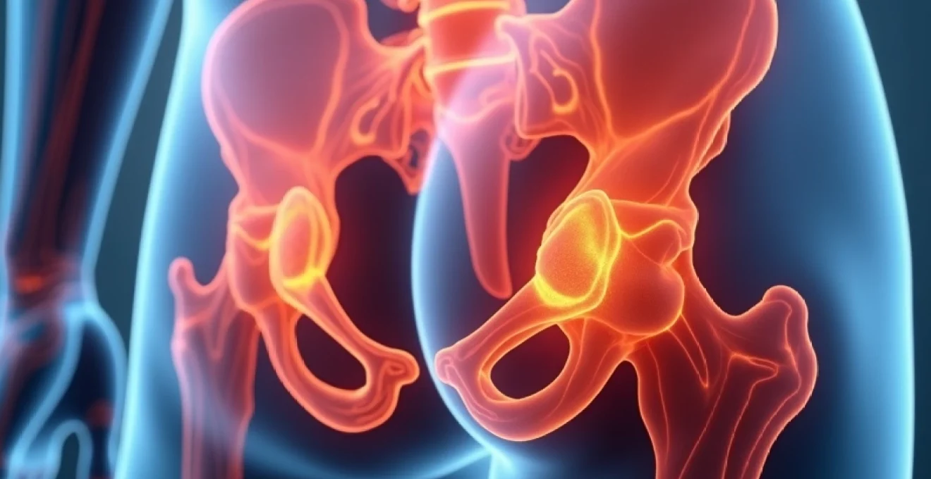
Anatomical connections between testicular and hip pain pathways
The intricate relationship between testicular and hip pain stems from fundamental anatomical connections established during embryonic development. The testicles originate from the same embryological tissue as the kidneys, positioned initially near the developing spine before descending into the scrotum during foetal development. This descent creates lasting neurological connections that explain why testicular pathology often manifests as referred pain in the hip, groin, and lower abdomen regions.
Genitofemoral nerve distribution and referred pain mechanisms
The genitofemoral nerve, arising from the L1-L2 spinal segments, serves as a primary pathway for referred pain between the testicular and hip regions. This nerve divides into genital and femoral branches, with the genital branch supplying the cremaster muscle and scrotal skin, whilst the femoral branch provides sensation to the upper thigh and hip area. When testicular pathology irritates the genital branch, pain signals can be perceived along the entire nerve distribution, creating the sensation of hip discomfort.
This referred pain mechanism operates through convergence of sensory fibres at the spinal cord level, where neurons receiving input from both testicular and hip regions may become cross-activated. The phenomenon explains why patients frequently describe deep, aching sensations that seem to migrate between the testicle and hip, particularly during movement or position changes that alter nerve tension.
Shared lymphatic drainage via para-aortic and iliac nodes
The lymphatic drainage patterns of both testicles and hip structures create another anatomical basis for associated pain syndromes. Testicular lymphatics drain primarily to the para-aortic and pre-aortic lymph nodes at the level of the renal vessels, whilst hip joint structures drain through the iliac and inguinal lymph node chains. When inflammatory conditions affect either region, the shared drainage pathways can propagate symptoms bidirectionally.
This lymphatic connection becomes particularly relevant in cases of testicular malignancy or infection, where enlarged lymph nodes may compress adjacent structures and create secondary hip pain. Similarly, hip infections or inflammatory conditions can affect the regional lymphatic flow, potentially creating testicular discomfort through retrograde inflammatory mediator spread.
L1-L2 spinal nerve root convergence patterns
The L1-L2 spinal nerve roots represent a critical convergence zone for sensory input from both testicular and hip regions. These nerve roots receive afferent fibres from testicular structures via the sympathetic nervous system, whilst simultaneously processing sensory information from hip joint capsule, surrounding musculature, and overlying skin. This convergence creates opportunities for cross-sensitisation and referred pain phenomena.
Spinal pathology affecting the L1-L2 segments, such as disc herniation or facet joint arthritis, can simultaneously affect both testicular and hip sensation. The resulting pain pattern often confuses diagnostic efforts, as patients may experience symptoms in both regions without clear localising features to guide clinical assessment.
Cremaster muscle innervation and hip flexor interactions
The cremaster muscle, responsible for testicular elevation and temperature regulation, shares fascial and neurological connections with the hip flexor muscle group. Both structures receive innervation from the genitofemoral nerve and maintain mechanical relationships through fascial continuity. When hip flexor pathology creates tension or inflammation, these forces can be transmitted to the cremaster muscle, resulting in testicular pain and altered testicular position.
Conversely, testicular pathology causing cremaster muscle spasm can create referred tension in the hip flexor group, manifesting as hip pain or restricted hip mobility. This biomechanical relationship explains why some patients report improved testicular symptoms following hip flexor stretching or massage therapy.
Testicular pathology causing ipsilateral hip pain
Primary testicular disorders frequently generate referred pain patterns that extend to the ipsilateral hip region. This phenomenon occurs through the previously described neurological pathways and represents a significant diagnostic consideration when evaluating patients with combined symptoms. Understanding these patterns enables clinicians to recognise when testicular pathology may be the underlying cause of apparent hip complaints.
Epididymitis and chronic pelvic pain syndrome manifestations
Epididymitis, characterised by inflammation of the coiled tube adjacent to the testicle, commonly produces referred pain that extends to the hip and groin regions. Acute bacterial epididymitis typically presents with severe testicular pain that radiates along the genitofemoral nerve distribution, creating deep aching sensations in the ipsilateral hip. Patients often describe difficulty walking or climbing stairs due to the combined testicular and hip discomfort.
Chronic epididymitis and associated chronic pelvic pain syndrome create more complex symptom patterns that can persist for months or years. The chronic inflammation leads to sensitisation of regional nerve pathways, resulting in persistent hip pain that may fluctuate with testicular symptoms. This condition frequently affects younger men and can significantly impact quality of life through its persistent nature and bilateral symptom distribution.
Testicular torsion emergency presentation with hip discomfort
Testicular torsion represents a urological emergency that frequently presents with referred hip pain alongside the characteristic testicular symptoms. The sudden onset of severe testicular pain from compromised blood supply triggers intense sympathetic nervous system activation, creating referred pain patterns that extend to the lower abdomen, groin, and hip. Patients typically present with severe pain that prevents normal ambulation and may initially attribute their discomfort to hip or groin injury.
The referred hip pain in testicular torsion often leads to diagnostic delays, particularly in younger patients who may not immediately recognise the testicular component of their symptoms. Rapid recognition of this pain pattern becomes crucial, as testicular salvage rates decline significantly beyond six hours from symptom onset. The combination of severe hip pain with nausea, vomiting, and altered testicular position should prompt immediate urological evaluation.
Varicocele-related venous congestion and referred pain
Varicoceles, representing dilated veins within the pampiniform plexus, create chronic testicular discomfort that frequently radiates to the hip region. The venous congestion leads to chronic hypoxia and inflammatory mediator accumulation, which sensitises regional pain pathways. Patients typically describe a dull, aching sensation that worsens with prolonged standing or physical activity and may extend from the testicle to the ipsilateral hip and lower abdomen.
The referred pain from varicoceles often follows the distribution of the testicular veins and associated lymphatic drainage. This creates characteristic pain patterns that may be mistaken for hip flexor strain or inguinal hernia, particularly in athletic populations where these conditions are common. The pain typically improves with recumbent positioning, which reduces venous congestion and associated inflammatory responses.
Orchitis secondary to mumps or bacterial infections
Testicular inflammation from viral or bacterial sources creates intense local pain that commonly refers to the hip through established neurological pathways. Mumps orchitis, typically affecting post-pubertal males, produces severe testicular swelling and pain that often radiates to the ipsilateral hip and lower abdomen. The inflammatory process triggers significant sympathetic nervous system activation, creating widespread referred pain patterns that may overshadow the primary testicular symptoms.
Bacterial orchitis, whether secondary to epididymitis or haematogenous spread, similarly produces referred hip pain through inflammatory mediator release and nerve sensitisation. The systemic inflammatory response often creates additional symptoms including fever, malaise, and regional lymphadenopathy, which can further complicate the diagnostic picture when hip pain predominates the clinical presentation.
Musculoskeletal hip disorders radiating to testicular region
Hip pathology frequently generates referred pain patterns that extend to the testicular region through shared innervation pathways and fascial connections. These conditions represent important differential diagnoses when evaluating patients with combined testicular and hip symptoms, as the primary pathology may actually originate from musculoskeletal structures rather than testicular abnormalities.
Femoroacetabular impingement and groin pain patterns
Femoroacetabular impingement (FAI) creates characteristic groin pain patterns that frequently extend to the testicular region through shared sensory innervation. The mechanical impingement between the femoral head and acetabulum triggers inflammatory responses that sensitise regional pain pathways, creating referred sensations that may be perceived as testicular discomfort. Young athletes particularly develop this condition due to repetitive hip flexion activities that gradually worsen the underlying bony abnormalities.
The referred testicular pain from FAI typically follows specific movement patterns, worsening with hip flexion, internal rotation, and adduction. This creates characteristic pain during activities such as getting into cars, climbing stairs, or sitting for prolonged periods. The pain pattern often confuses diagnostic efforts when patients focus primarily on the testicular component of their symptoms rather than the underlying hip pathology.
Iliopsoas bursitis with inguinal canal compression
Inflammation of the iliopsoas bursa creates deep groin pain that can extend to the testicular region through mechanical compression of the inguinal canal structures. The inflamed bursa, located between the iliopsoas tendon and the hip joint capsule, can become enlarged and compress adjacent structures including the lateral femoral cutaneous nerve and components of the inguinal canal. This compression creates referred pain patterns that may be perceived as testicular discomfort.
Patients with iliopsoas bursitis typically describe deep, aching pain that worsens with hip flexion and improves with hip extension. The referred testicular component often fluctuates with hip position and activity level, providing important diagnostic clues that distinguish this condition from primary testicular pathology. Physical examination revealing tenderness over the iliopsoas tendon and reproduction of symptoms with hip flexion against resistance supports this diagnosis.
Avascular necrosis of femoral head pain distribution
Avascular necrosis of the femoral head produces deep, constant hip pain that frequently refers to the groin and testicular regions through the obturator nerve pathway. The progressive bone death creates inflammatory responses that sensitise regional pain pathways, resulting in referred pain patterns that may initially be mistaken for testicular pathology. This condition particularly affects younger men with risk factors such as corticosteroid use, alcohol consumption, or previous hip trauma.
The referred testicular pain from avascular necrosis typically presents as a deep, aching sensation that persists at rest and worsens with weight-bearing activities. Unlike primary testicular pathology, the pain often improves with non-weight-bearing positions and may be associated with progressive hip stiffness and functional limitations. Early recognition becomes crucial as the condition may be reversible with appropriate intervention before femoral head collapse occurs.
Piriformis syndrome and sciatic nerve involvement
Piriformis syndrome, characterised by compression or irritation of the sciatic nerve by the piriformis muscle, can create complex referred pain patterns that include testicular discomfort. The sciatic nerve shares spinal cord segments with testicular innervation, creating opportunities for cross-sensitisation and referred pain phenomena. Athletes and individuals with prolonged sitting occupations particularly develop this condition due to repetitive piriformis muscle overuse.
The testicular component of piriformis syndrome typically manifests as a deep, aching sensation that worsens with prolonged sitting or hip rotation activities. Patients often describe difficulty pinpointing the exact source of their discomfort, as the pain may seem to migrate between the hip, buttock, and testicular regions. Physical examination techniques that stress the piriformis muscle can help reproduce symptoms and confirm this diagnosis.
Urological conditions presenting with combined symptoms
Several urological conditions create symptom patterns that simultaneously affect both testicular and hip regions, representing important diagnostic considerations when evaluating patients with this complaint pattern. These conditions often involve structures that anatomically bridge the gap between the urogenital and musculoskeletal systems, creating complex symptom presentations that require comprehensive urological evaluation.
Inguinal hernias represent one of the most common urological conditions causing combined testicular and hip symptoms. When abdominal contents protrude through the inguinal canal, they can compress testicular blood supply and innervation whilst simultaneously creating hip pain through mechanical effects on surrounding musculature. Large indirect inguinal hernias may extend into the scrotum, creating obvious testicular symptoms, whilst smaller hernias may produce subtle testicular discomfort alongside hip pain that worsens with increased intra-abdominal pressure.
Kidney stones affecting the right ureter frequently create referred pain patterns that involve both testicular and hip regions through shared visceral innervation pathways. The intense pain from ureteral spasm triggers widespread sympathetic nervous system activation, creating referred sensations that extend far beyond the primary urological pathology. Patients typically describe severe, colicky pain that radiates from the flank to the groin, testicle, and hip, often accompanied by nausea, vomiting, and urinary symptoms.
Chronic prostatitis and chronic pelvic pain syndrome create persistent symptoms that may involve both testicular and hip regions through inflammatory mediator effects on regional nerve pathways. The chronic inflammation sensitises pelvic floor muscles and associated fascial connections, creating referred pain patterns that can extend to the hip flexor group and testicular region. This condition particularly affects younger men and may persist for months or years despite various treatment approaches.
Urinary tract infections affecting the bladder or prostate can create referred pain patterns that involve the testicular and hip regions through sympathetic nervous system activation and inflammatory mediator release. The infection triggers local inflammatory responses that sensitise regional pain pathways, creating referred sensations that may be perceived as testicular or hip discomfort. Systemic symptoms such as fever, malaise, and urinary symptoms typically accompany these conditions, providing important diagnostic clues.
Diagnostic imaging protocols for Right-Sided pain assessment
Comprehensive imaging evaluation of patients presenting with right testicular and hip pain requires a systematic approach that addresses both potential primary pathologies and referred pain mechanisms. The imaging protocol must be tailored to the specific clinical presentation whilst maintaining cost-effectiveness and minimising unnecessary radiation exposure. Modern imaging techniques provide unprecedented detail of both testicular and hip structures, enabling accurate diagnosis of conditions that previously required invasive diagnostic procedures.
Scrotal ultrasonography serves as the primary imaging modality for evaluating testicular pathology in patients with combined symptoms. High-resolution ultrasound with colour Doppler capability can assess testicular blood flow, identify masses or inflammatory changes, and evaluate the epididymis and surrounding structures. The examination should include assessment of both testicles for comparison, measurement of testicular volumes, and evaluation of the pampiniform plexus for varicocele identification. When testicular torsion is suspected, immediate ultrasound evaluation becomes crucial for determining testicular viability and guiding emergency intervention.
Hip imaging typically begins with plain radiographs to assess bony anatomy and identify obvious pathology such as fractures, arthritis, or avascular necrosis. Standard views include anteroposterior pelvis and lateral hip projections, which can identify most significant bony abnormalities. However, advanced imaging techniques such as magnetic resonance imaging (MRI) are often necessary to evaluate soft tissue structures, assess for labral tears or impingement, and identify early avascular necrosis changes that may not be apparent on plain films.
Cross-sectional imaging with computed tomography (CT) or MRI may be indicated when clinical presentation suggests referred pain from retroperitoneal or intra-abdominal pathology. CT urography can identify kidney stones, ureteral obstructions, or other urological pathology that may present with combined testicular and hip symptoms. MRI provides superior soft tissue contrast and can identify inflammatory conditions, mass lesions, or vascular abnormalities that may contribute to the symptom complex.
Specialised imaging techniques such as magnetic resonance arthrography of the hip may be necessary to evaluate suspected labral tears or cartilage abnormalities in patients with suspecte
d femoroacetabular impingement or other intra-articular pathology. Nuclear medicine studies, such as bone scintigraphy, may occasionally be useful for identifying occult fractures or inflammatory conditions affecting the hip region when conventional imaging remains inconclusive.
The imaging sequence should be guided by clinical presentation and physical examination findings. Patients with obvious testicular abnormalities should undergo scrotal ultrasound as the initial study, whilst those with predominantly hip symptoms may benefit from hip-specific imaging first. Systematic evaluation protocols that address both potential pathologies ensure comprehensive assessment whilst avoiding unnecessary duplicate studies or delayed diagnosis.
Conservative and surgical treatment approaches by underlying aetiology
Treatment strategies for patients with right testicular and hip pain must address the underlying pathology whilst managing the complex symptom interactions that characterise this presentation. Conservative approaches often provide significant symptom relief for many conditions, whilst surgical intervention becomes necessary for specific pathologies that threaten organ viability or create progressive functional impairment. The treatment approach should be individualised based on accurate diagnosis, symptom severity, and patient-specific factors including age, activity level, and comorbid conditions.
Conservative management for testicular pathology typically includes anti-inflammatory medications, scrotal support, activity modification, and physical therapy targeting pelvic floor dysfunction when appropriate. Acute epididymitis responds well to antibiotic therapy combined with scrotal elevation and ice application, whilst chronic epididymitis may require prolonged courses of anti-inflammatory medications and neuropathic pain agents. Varicocele-related symptoms often improve with scrotal support garments and avoidance of prolonged standing, though surgical intervention may be necessary for persistent symptoms or fertility concerns.
Hip-related pathology requires treatment strategies targeting the specific underlying condition. Femoroacetabular impingement often responds to physical therapy focusing on hip mobility and strengthening exercises, combined with activity modification to avoid impingement positions. When conservative measures fail, arthroscopic hip surgery can address labral tears and bony impingement. Iliopsoas bursitis typically responds to targeted injection therapy and stretching exercises, whilst avascular necrosis may require surgical intervention ranging from core decompression to hip replacement depending on disease stage.
Surgical intervention for testicular pathology includes orchidopexy for testicular torsion, varicocelectomy for symptomatic varicoceles, and orchiectomy for testicular malignancy or irreversible torsion. Emergency surgical exploration remains the gold standard for suspected testicular torsion, as clinical examination alone cannot reliably exclude this diagnosis. Prompt surgical intervention within six hours of symptom onset provides the best opportunity for testicular salvage, emphasising the importance of rapid diagnosis and treatment.
Combined treatment approaches often provide optimal outcomes for patients with complex symptom patterns involving both testicular and hip regions. Multidisciplinary care involving urologists, orthopaedic surgeons, and pain management specialists can address the various components of these challenging presentations. Physical therapy targeting both pelvic floor dysfunction and hip biomechanics often provides significant symptom improvement, particularly for patients with chronic pain syndromes affecting both regions.
Pain management strategies must consider the complex neurological interactions between testicular and hip regions. Neuropathic pain medications such as gabapentin or pregabalin can be effective for chronic pain syndromes involving nerve sensitisation, whilst targeted injection therapy may provide relief for specific anatomical sources of pain. Regional anaesthetic blocks of the genitofemoral nerve or lumbar sympathetic chain can provide both diagnostic information and therapeutic benefit for patients with complex referred pain patterns.
Follow-up care requires ongoing assessment of both symptom resolution and functional improvement. Patients should be monitored for recurrence of testicular pathology, progression of hip disorders, and development of chronic pain syndromes that may require long-term management strategies. Regular reassessment ensures appropriate treatment modifications and early identification of complications that may require additional intervention. The complex nature of combined testicular and hip symptoms often requires patience and persistence from both patients and healthcare providers to achieve optimal outcomes.