Sharp pain in the right upper back can transform routine activities into challenging ordeals, affecting everything from breathing deeply to reaching for objects overhead. This distinctive type of discomfort often manifests as sudden, stabbing sensations that can radiate across the shoulder blade, between the ribs, or toward the neck and arm. Unlike the dull ache commonly associated with general back tension, sharp upper back pain typically indicates specific anatomical structures under stress or dysfunction.
The complexity of the upper back’s anatomy means that multiple systems can contribute to sharp, localised pain on the right side. From intricate muscle networks that support shoulder blade movement to the thoracic spine’s unique relationship with the rib cage, various structures can generate intense, focused discomfort. Understanding these potential sources becomes crucial for anyone experiencing persistent or recurring episodes of sharp right upper back pain.
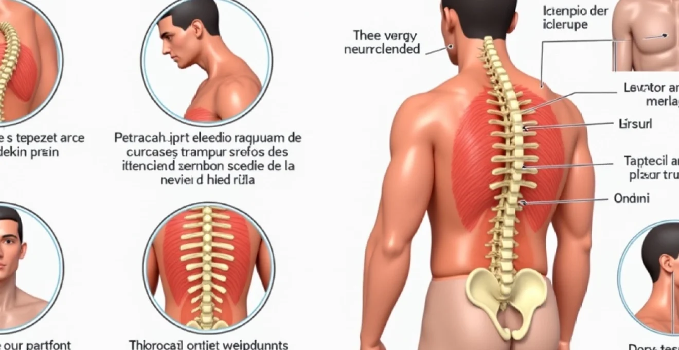
Musculoskeletal origins of right upper back sharp pain
The musculoskeletal system represents the most common source of sharp right upper back pain, with various muscles, tendons, and fascial structures capable of producing intense, localised discomfort. These tissues work in complex coordination to support posture, facilitate arm movement, and maintain spinal stability. When dysfunction occurs within this intricate network, the resulting pain often presents as sharp, stabbing sensations that can severely impact daily function.
Rhomboid muscle strain and trigger point formation
The rhomboid muscles, consisting of the rhomboid major and minor, play a crucial role in retracting the shoulder blade toward the spine. These diamond-shaped muscles frequently develop strain patterns and trigger points that manifest as sharp, burning pain between the shoulder blade and spine. Trigger points within the rhomboids typically create referred pain patterns that can extend from the medial border of the scapula toward the posterior shoulder region.
Rhomboid dysfunction commonly develops from prolonged forward head posture, repetitive reaching activities, or sudden overhead movements that exceed the muscle’s capacity. The pain characteristics often include sharp stabbing sensations when moving the arm across the body or when attempting to squeeze the shoulder blades together. Additionally, individuals may experience a constant ache that intensifies with sustained positions or specific movement patterns.
Trapezius muscle spasm in the middle fibres
The middle fibres of the trapezius muscle are particularly susceptible to spasm and trigger point development, creating sharp pain that radiates across the upper back region. These muscle fibres are responsible for retracting the scapula and supporting the weight of the arms during various activities. When overloaded or fatigued, they can develop protective spasms that generate intense, localised pain.
Trapezius spasms often result from poor ergonomic positioning, emotional stress, or sudden movements that overwhelm the muscle’s capacity. The pain typically presents as a sharp, gripping sensation that may be accompanied by visible muscle knots or bands. Individuals frequently report difficulty sleeping on the affected side and increased pain with neck rotation or shoulder elevation movements.
Levator scapulae muscle tension and cervical dysfunction
The levator scapulae muscle connects the upper cervical vertebrae to the superior angle of the scapula, making it vulnerable to both neck and shoulder blade dysfunction. This muscle commonly develops chronic tension patterns that can produce sharp, radiating pain from the neck down to the upper back region. The unique anatomical position of this muscle means that dysfunction can create pain that seems to originate from multiple sources simultaneously.
Levator scapulae tension frequently develops from sustained neck flexion, such as prolonged computer work or reading in poor positions. The resulting pain often presents as a sharp, aching sensation that follows the muscle’s pathway from the neck to the shoulder blade. Individuals may also experience associated symptoms including neck stiffness, headaches, and difficulty with overhead arm movements.
Serratus anterior weakness and scapular dyskinesis
The serratus anterior muscle plays a vital role in stabilising the shoulder blade against the rib cage and facilitating proper arm elevation mechanics. When this muscle becomes weak or inhibited, compensatory patterns develop in surrounding muscles, potentially leading to sharp upper back pain. Scapular dyskinesis , or abnormal shoulder blade movement, often results from serratus anterior dysfunction and can create secondary pain patterns.
Serratus anterior weakness commonly manifests as sharp pain during overhead activities or when attempting to push objects away from the body. The compensation patterns that develop can overload the rhomboids, middle trapezius, and other scapular stabilisers, creating a cascade of dysfunction that generates persistent sharp pain in the upper back region.
Intercostal muscle strain between T3-T6 vertebrae
The intercostal muscles, located between adjacent ribs, can develop strain patterns that create sharp, stabbing pain along the rib cage and into the upper back. These muscles are essential for respiratory function and trunk rotation, making them susceptible to injury during twisting movements or forced breathing patterns. Intercostal muscle strain between the T3-T6 levels commonly refers pain toward the posterior chest and upper back region.
Intercostal dysfunction often develops from sudden rotational movements, prolonged coughing episodes, or direct trauma to the chest wall. The resulting pain typically presents as sharp, knife-like sensations that worsen with deep breathing, coughing, or trunk rotation. Individuals may also experience muscle spasms that create visible chest wall asymmetries and further compromise respiratory function.
Thoracic spine pathology contributing to sharp upper back pain
The thoracic spine’s unique anatomy, with its connection to the rib cage and relatively limited mobility compared to the cervical and lumbar regions, creates specific pathological patterns that can generate sharp upper back pain. Unlike other spinal regions, the thoracic spine must balance stability requirements for organ protection with sufficient mobility for respiratory function and arm movement. This delicate balance makes certain structures particularly vulnerable to dysfunction and subsequent pain generation.
T4-T5 facet joint dysfunction and capsular restriction
The facet joints of the mid-thoracic spine, particularly at the T4-T5 level, are prone to developing dysfunction that creates sharp, localised pain. These small joints guide spinal movement and can become restricted due to poor posture, repetitive stress, or minor trauma. When facet joint capsules become inflamed or restricted, they generate sharp pain that often radiates along the rib cage or toward the shoulder blade region.
T4-T5 facet joint dysfunction commonly presents as sharp pain that worsens with spinal extension or rotation toward the affected side. The pain may be accompanied by a sensation of joint “catching” or “locking” during certain movements. Capsular restriction at these levels can also create compensatory movement patterns that overload adjacent spinal segments and perpetuate the pain cycle.
Thoracic disc herniation at T3-T4 and T5-T6 levels
While less common than cervical or lumbar disc herniations, thoracic disc problems can create intense sharp pain that radiates around the rib cage or into the upper back. The T3-T4 and T5-T6 levels are particularly susceptible to disc degeneration and herniation due to their location at the apex of the thoracic kyphosis. When disc material protrudes or extrudes, it can compress neural structures and create sharp, radiating pain patterns.
Thoracic disc herniation often presents as sharp, burning pain that follows a dermatomal pattern around the chest wall or into the upper back. The pain may worsen with coughing, sneezing, or Valsalva manoeuvres that increase intrathecal pressure. In severe cases, individuals may experience numbness or tingling that follows the affected nerve root distribution, creating a band-like sensation around the chest.
Costotransverse joint irritation and rib subluxation
The costotransverse joints, where the ribs articulate with the thoracic vertebrae, can develop dysfunction that creates sharp, localised pain in the upper back region. These joints are essential for respiratory mechanics and can become irritated through repetitive stress, trauma, or inflammatory conditions. When costotransverse joint dysfunction occurs, it often creates sharp pain that worsens with breathing or trunk rotation.
Rib subluxation , though controversial in its exact mechanism, describes a condition where normal rib mechanics become altered, creating pain and dysfunction. This condition commonly affects the upper ribs and can generate sharp pain that radiates from the spine toward the anterior chest wall. The pain typically worsens with deep inspiration or specific rotational movements that stress the affected joint.
Scheuermann’s disease and thoracic kyphosis complications
Scheuermann’s disease, a developmental condition affecting the thoracic spine during adolescence, can create long-term complications that manifest as sharp upper back pain in adulthood. This condition involves irregular vertebral development that creates increased thoracic kyphosis and potential instability at the apex of the curve. The resulting biomechanical alterations can predispose individuals to various pain syndromes throughout their adult lives.
Adults with a history of Scheuermann’s disease often experience sharp pain at the apex of their thoracic curve, typically around the T6-T8 levels. The pain may result from accelerated facet joint degeneration, disc degeneration, or muscle imbalances created by the altered spinal alignment. These individuals are also more susceptible to developing secondary conditions such as costotransverse joint dysfunction or intercostal neuralgia.
Nerve-related causes of Right-Sided upper back sharp pain
Neurological sources of sharp right upper back pain involve compression, irritation, or dysfunction of specific nerve structures that innervate the upper back region. These conditions often create characteristic pain patterns that follow anatomical nerve distributions, helping clinicians identify the specific neural structure involved. The complexity of nerve pathways in the upper back means that dysfunction can occur at multiple levels, from nerve roots exiting the spine to peripheral nerve branches that innervate muscles and skin.
Thoracic outlet syndrome and brachial plexus compression
Thoracic outlet syndrome encompasses a group of conditions involving compression of neurovascular structures as they traverse the space between the clavicle and first rib. While commonly associated with arm symptoms, thoracic outlet syndrome can create sharp pain that radiates into the upper back, particularly when the brachial plexus nerves are involved. The neurogenic variant of this condition often produces sharp, shooting pain that follows specific nerve distributions.
Brachial plexus compression within the thoracic outlet can create pain patterns that extend from the neck into the upper back, shoulder, and arm regions. The pain typically worsens with overhead arm positions or activities that narrow the thoracic outlet space. Individuals may also experience associated symptoms including numbness, tingling, and weakness that follow specific nerve root distributions.
Intercostal neuralgia following T4-T6 nerve distribution
Intercostal neuralgia involves irritation or inflammation of the intercostal nerves that travel along the undersurface of the ribs. When nerves at the T4-T6 levels are affected, the resulting pain often radiates from the spine around the rib cage and can create sharp, burning sensations in the upper back region. This condition can develop following viral infections, trauma, or surgical procedures that affect the intercostal nerve pathways.
The pain associated with intercostal neuralgia typically follows a dermatomal distribution, creating a band-like pattern of sharp, shooting pain. Individuals often describe the sensation as electric shock-like pain that can be triggered by light touch, movement, or changes in position. The condition may also create allodynia, where normally non-painful stimuli become painful, significantly impacting daily activities and quality of life.
Dorsal scapular nerve entrapment and C5 root irritation
The dorsal scapular nerve, arising from the C5 nerve root, innervates the rhomboid muscles and part of the middle trapezius. Entrapment of this nerve can occur as it pierces the middle scalene muscle or becomes compressed by other anatomical structures. When compromised, the dorsal scapular nerve creates sharp pain between the shoulder blades that may be accompanied by muscle weakness and altered scapular mechanics.
C5 nerve root irritation can also create referred pain patterns that extend into the upper back region, particularly along the distribution of the dorsal scapular nerve. This condition often develops from cervical spine dysfunction, disc herniation, or foraminal stenosis that compresses the nerve root as it exits the spine. The resulting pain may be accompanied by weakness in rhomboid function and altered shoulder blade movement patterns.
Long thoracic nerve palsy and serratus anterior denervation
The long thoracic nerve innervates the serratus anterior muscle and can become compromised through various mechanisms including trauma, repetitive overhead activities, or viral infections. When this nerve becomes damaged or compressed, it creates characteristic weakness in the serratus anterior muscle, leading to scapular winging and compensatory pain patterns in the upper back region.
Long thoracic nerve palsy often creates sharp pain that develops secondarily to the muscle imbalances caused by serratus anterior weakness. As other muscles compensate for the lost function, they become overloaded and develop trigger points or strain patterns that generate sharp upper back pain. The condition may also create direct nerve-related pain that follows the anatomical distribution of the long thoracic nerve.
Referred pain patterns manifesting as right upper back sharpness
Referred pain represents a fascinating phenomenon where dysfunction in one anatomical region creates pain perception in a distant area through complex neurological pathways. The upper back frequently serves as a target for referred pain from various internal organs, cervical spine structures, and even cardiac conditions. Understanding these referral patterns becomes crucial for clinicians and patients alike, as treating only the area where pain is felt may miss the actual source of dysfunction.
Cardiac conditions, particularly those affecting the right side of the heart or involving the pericardium, can create referred pain that manifests as sharp sensations in the right upper back. While left-sided chest pain remains the classic presentation for cardiac events, right-sided cardiac issues or atypical presentations can generate pain that radiates to the right shoulder blade and upper back region. Coronary artery spasms or right ventricular strain can create sharp, stabbing pain that mimics musculoskeletal conditions.
Gallbladder dysfunction represents another common source of referred pain to the right upper back region. Cholecystitis, choledocholithiasis, or gallbladder inflammation can create sharp pain that radiates from the right upper quadrant of the abdomen to the right shoulder blade and upper back. This pain often accompanies digestive symptoms but may occasionally present as isolated upper back pain, particularly in atypical cases or when gallbladder dysfunction develops gradually.
Pulmonary conditions affecting the right lung can also generate referred pain patterns that manifest as sharp upper back discomfort. Pleuritis, pneumonia, or pulmonary embolism can create pain that radiates to the posterior chest wall and upper back region. The pain typically worsens with deep inspiration or coughing and may be accompanied by respiratory symptoms, though these may be subtle in early stages of lung pathology.
Cervical spine dysfunction frequently refers pain into the upper back region through complex neurological pathways. Facet joint dysfunction at the C6-C7 or C7-T1 levels can create sharp pain that radiates into the upper trapezius and rhomboid regions. Similarly, cervical disc herniations or nerve root compressions can generate pain patterns that extend well beyond the neck into the upper back and shoulder blade areas.
The intricate connections between cervical nerve roots and upper back musculature mean that neck dysfunction can create persistent upper back pain that fails to respond to local treatment approaches.
Diagnostic assessment techniques for right upper back sharp pain
Accurate diagnosis of sharp right upper back pain requires a systematic approach that considers the complex anatomy and multiple potential pain generators in this region. The diagnostic process must differentiate between local musculoskeletal dysfunction, referred pain patterns, and potentially serious underlying conditions that may require immediate medical attention. A comprehensive assessment combines detailed history taking, physical examination techniques, and appropriate imaging studies to identify the primary pain source.
The clinical history provides crucial insights into the nature and origin of sharp upper back pain. Pain onset patterns help distinguish between acute traumatic injuries and gradual onset conditions related to postural stress or degenerative changes. Sharp pain that develops suddenly following a specific incident suggests acute muscle strain, joint dysfunction, or even more serious conditions requiring immediate evaluation. Conversely, pain that develops gradually over weeks or months
typically points toward chronic conditions such as postural dysfunction, muscle imbalances, or degenerative joint changes.The location and radiation patterns of pain provide valuable diagnostic clues about the underlying structure involved. Sharp pain that remains localised to specific muscle groups suggests primary musculoskeletal dysfunction, while pain that radiates in dermatomal patterns indicates potential nerve involvement. Pain quality descriptors help differentiate between muscle-related pain (often described as aching or cramping), joint-related pain (typically sharp and localised), and nerve-related pain (frequently described as burning, shooting, or electric-like sensations).Physical examination techniques must systematically assess all potential pain generators in the upper back region. Postural analysis reveals alignment abnormalities that may contribute to muscle imbalances and joint dysfunction. Palpation of specific muscle groups, including the rhomboids, trapezius, and intercostal muscles, can identify trigger points, muscle knots, or areas of heightened sensitivity that correspond to the patient’s pain complaints.Range of motion testing provides insights into joint mobility restrictions and movement patterns that reproduce or alleviate symptoms. Active and passive movements of the neck, shoulders, and thoracic spine help identify specific structures involved in pain generation. Provocative tests such as the foraminal compression test, thoracic outlet manoeuvres, and specific muscle stretch tests can reproduce symptoms and confirm suspected diagnoses.Imaging studies play a supportive role when clinical findings suggest structural abnormalities or when conservative treatment approaches fail to provide adequate relief. Plain radiographs can reveal gross structural abnormalities, vertebral alignment issues, or signs of degenerative changes. Magnetic resonance imaging provides detailed visualisation of soft tissue structures, including muscles, discs, and neural elements, making it particularly valuable for suspected disc herniations or nerve compressions.Advanced diagnostic techniques such as diagnostic nerve blocks or trigger point injections can serve both therapeutic and diagnostic purposes. These procedures help confirm the specific anatomical source of pain while providing temporary relief that validates the treatment approach. Electromyographic studies may be indicated when nerve dysfunction is suspected, helping to localise the level and extent of neural involvement.
Conservative treatment protocols for acute right upper back pain episodes
Conservative management represents the first-line approach for most cases of sharp right upper back pain, with evidence supporting the effectiveness of multimodal treatment strategies that address both symptom relief and underlying dysfunction. The treatment approach must be tailored to the specific pain source identified through diagnostic assessment, while also addressing contributing factors such as postural abnormalities, muscle imbalances, and lifestyle factors that perpetuate the condition.Immediate pain management during acute episodes focuses on reducing inflammation and muscle guarding that can perpetuate the pain cycle. Cold therapy applied within the first 48-72 hours helps reduce inflammation and provides analgesic effects through neurological mechanisms. Ice applications should be limited to 15-20 minute sessions every 2-3 hours to prevent tissue damage while maximising therapeutic benefits. After the acute inflammatory phase, heat therapy can help reduce muscle tension and improve tissue extensibility, preparing tissues for therapeutic interventions.Pharmacological interventions support the healing process while providing symptomatic relief during acute pain episodes. Nonsteroidal anti-inflammatory drugs (NSAIDs) address both pain and inflammation, though they should be used judiciously and with consideration of individual contraindications. Muscle relaxants may be beneficial for cases involving significant muscle spasm, though their use should be time-limited to prevent dependency. Topical analgesics provide localised pain relief without systemic side effects and can be particularly effective for superficial musculoskeletal pain sources.Manual therapy techniques form a cornerstone of conservative treatment for upper back pain, with various approaches targeting different aspects of dysfunction. Soft tissue mobilisation techniques, including massage and myofascial release, help reduce muscle tension and improve tissue quality. Joint mobilisation and manipulation techniques can restore normal movement patterns and reduce pain associated with joint restrictions. Trigger point therapy specifically targets hyperactive areas within muscles that contribute to localised and referred pain patterns.Therapeutic exercise programs must address both immediate symptom relief and long-term prevention of recurrence. Stretching exercises focus on improving flexibility in commonly tight muscle groups, including the upper trapezius, levator scapulae, and pectoral muscles that contribute to forward head posture. Strengthening exercises target weak or inhibited muscles such as the deep neck flexors, rhomboids, and middle trapezius that support optimal postural alignment.Progressive loading strategies help tissues adapt to increasing demands while preventing re-injury during the recovery process. Initial exercises may focus on gentle range of motion and basic activation of key muscle groups. As symptoms improve, exercises progress to include resistance training, functional movement patterns, and eventually sport-specific or occupation-specific activities that prepare individuals for return to full activity levels.Postural education and ergonomic modifications address underlying factors that contribute to upper back pain development and recurrence. Workplace assessments can identify problematic positions or equipment that stress the upper back region. Ergonomic adjustments such as monitor positioning, chair height, and keyboard placement can significantly reduce postural stress on the neck and upper back during prolonged sitting activities.Sleep positioning education helps optimise recovery during rest periods and prevents positions that perpetuate muscle tension or joint restriction. Pillow selection and mattress considerations play important roles in maintaining neutral spinal alignment during sleep. Individuals may benefit from specific positioning strategies that reduce stress on healing tissues while promoting optimal recovery conditions.Activity modification strategies help individuals maintain function while avoiding activities that exacerbate symptoms during the acute recovery phase. This approach involves identifying specific movements or positions that trigger pain and developing alternative strategies to accomplish necessary tasks. Temporary activity restrictions should be balanced with the need to maintain general fitness and prevent deconditioning that can complicate recovery.Stress management techniques address the psychological and emotional factors that can influence pain perception and muscle tension. Relaxation techniques, breathing exercises, and mindfulness practices can help reduce muscle guarding and improve pain tolerance. Stress reduction strategies may also help address underlying tension patterns that contribute to chronic muscle dysfunction in the upper back region.Patient education throughout the treatment process empowers individuals to take an active role in their recovery and helps prevent future episodes of upper back pain. Understanding the relationship between posture, movement patterns, and pain development enables patients to make informed decisions about daily activities and self-care strategies. Education about warning signs that indicate the need for professional intervention helps patients seek appropriate care before minor issues progress to more significant problems.Progressive return to activity protocols ensure safe resumption of full function while minimising the risk of re-injury or symptom recurrence. These protocols involve gradual increases in activity intensity and duration, with careful monitoring of symptom response. Successful progression through return-to-activity phases indicates tissue healing and restored functional capacity, while symptom recurrence suggests the need for modified progression or additional therapeutic intervention.