The human cervical spine naturally forms a gentle, inward-curving arc that plays a fundamental role in supporting the weight of the head whilst maintaining optimal biomechanical function. When this natural curvature becomes diminished or completely flattened, a condition known as cervical spine straightening or loss of cervical lordosis occurs. This anatomical alteration affects millions of individuals worldwide, particularly in our increasingly sedentary, technology-driven society where prolonged periods of poor posture have become commonplace.
Understanding the implications of cervical spine straightening extends far beyond mere postural concerns. The loss of natural cervical lordosis represents a significant departure from optimal spinal biomechanics, potentially triggering a cascade of compensatory mechanisms throughout the entire musculoskeletal system. From increased susceptibility to degenerative changes to compromised neurological function, the ramifications of this condition can be both immediate and long-lasting, making early recognition and appropriate intervention crucial for maintaining spinal health and preventing progressive deterioration.
Cervical lordosis anatomy and normal spinal curvature mechanics
The cervical spine comprises seven vertebrae designated C1 through C7, each meticulously designed to contribute to the overall structural integrity and functional capacity of the neck region. Under normal circumstances, these vertebrae align to form a gentle, inward-facing curve known as cervical lordosis, typically measuring between 20 and 40 degrees when assessed radiographically. This curvature represents one of the spine’s four natural curves, working in harmony with thoracic kyphosis, lumbar lordosis, and sacral kyphosis to create the characteristic S-shaped configuration that defines healthy spinal alignment.
The significance of maintaining proper cervical lordosis cannot be overstated, as this curvature serves multiple critical functions within the broader context of spinal biomechanics. The lordotic curve acts as a natural shock absorber , distributing mechanical forces evenly throughout the cervical region whilst reducing concentrated stress on individual vertebral segments. Additionally, this anatomical configuration optimises the positioning of neural structures, ensuring adequate space within the spinal canal for the cervical spinal cord and emerging nerve roots.
C-shaped lordotic curve function in weight distribution
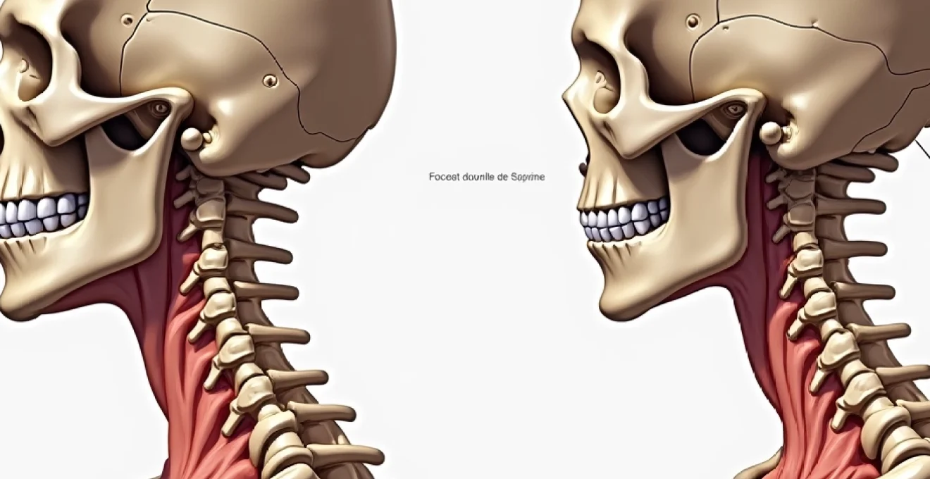
The natural C-shaped lordotic curve of the cervical spine operates as a sophisticated engineering marvel, efficiently managing the substantial load imposed by the human head, which typically weighs between 10 and 12 pounds. When positioned correctly above the shoulders, this weight is distributed evenly throughout the cervical vertebrae, minimising stress concentrations and reducing the risk of premature degenerative changes. However, when the cervical lordosis is lost or significantly diminished, the biomechanical advantages of this natural architecture are compromised.
Forward head posture , often accompanying cervical spine straightening, dramatically alters the mechanical loading patterns throughout the neck region. For every inch the head moves forward from its optimal position, the effective weight bearing down on the cervical spine increases exponentially, potentially doubling or even tripling the forces that must be managed by the surrounding musculature and skeletal structures.
Atlas-axis complex role in cervical alignment
The atlas-axis complex, comprising the first and second cervical vertebrae (C1 and C2), plays a pivotal role in maintaining overall cervical alignment and facilitating the intricate movements required for normal head and neck function. The atlas, or C1 vertebra, lacks a traditional vertebral body and instead forms a ring-like structure that supports the occiput of the skull. This unique anatomy allows for the nodding movements of the head whilst providing a stable platform for weight transmission.
The axis, or C2 vertebra, features the distinctive odontoid process (dens) that projects upward through the atlas, creating a sophisticated pivot joint that enables rotational movements of the head. When cervical spine straightening occurs, the normal relationships between these critical structures become altered, potentially compromising both stability and mobility throughout the upper cervical region. Dysfunction at the atlas-axis level can have far-reaching effects on the entire cervical spine, contributing to compensatory patterns that may exacerbate the loss of normal lordotic curvature.
Intervertebral disc height variations from C2-C7
The intervertebral discs of the cervical spine exhibit distinct height variations from C2-C7, with each disc contributing to the overall maintenance of cervical lordosis through its unique morphological characteristics. These fibrocartilaginous structures serve as both shock absorbers and spacers between adjacent vertebrae, with their wedge-shaped configuration playing a crucial role in creating and maintaining the natural cervical curve. The anterior (front) portion of each cervical disc is typically thicker than the posterior (back) portion, contributing approximately 60% to the overall lordotic angulation.
When cervical spine straightening occurs, the normal disc height relationships become compromised, often resulting in accelerated degenerative changes throughout the affected segments.
The loss of disc height anteriorly can significantly contribute to the flattening of cervical lordosis, creating a self-perpetuating cycle of progressive structural deterioration.
This process is particularly evident in the lower cervical segments (C5-C6 and C6-C7), where the greatest degree of flexion and extension movements typically occur.
Facet joint orientation and biomechanical load transfer
The facet joints of the cervical spine are uniquely oriented to facilitate the complex range of motions required for normal head and neck function whilst providing essential stability during weight-bearing activities. These synovial joints, positioned at approximately 45 degrees to the horizontal plane, allow for smooth gliding movements during flexion, extension, and lateral bending whilst limiting excessive translation between adjacent vertebrae. The orientation of cervical facet joints also plays a crucial role in maintaining normal lordotic curvature by providing posterior restraint against forward sliding of the vertebrae.
When cervical spine straightening occurs, the normal biomechanical load transfer patterns through the facet joints become significantly altered. The loss of natural lordosis can lead to increased compressive forces on the posterior elements of the cervical vertebrae, potentially resulting in facet joint arthritis, capsular irritation, and referred pain patterns. This mechanical dysfunction often contributes to the development of muscle spasm and further postural compensations that can exacerbate the underlying condition.
Pathophysiology of cervical spine straightening and loss of lordosis
The pathophysiology underlying cervical spine straightening involves a complex interplay of mechanical, biochemical, and neurological factors that collectively contribute to the progressive loss of normal lordotic curvature. Understanding these mechanisms is essential for developing effective treatment strategies and preventing further deterioration. The process typically begins with acute or chronic stressors that disrupt normal spinal mechanics, leading to compensatory adaptations that may initially provide short-term relief but ultimately result in long-term structural changes.
Multiple factors can initiate the cascade of events leading to cervical spine straightening, including traumatic injuries, prolonged postural stress, degenerative changes, and inflammatory conditions. The body’s attempt to protect injured or stressed tissues often involves muscle guarding and protective positioning, which can temporarily alter normal spinal alignment. However, when these protective mechanisms persist over extended periods, they can lead to permanent structural adaptations that compromise optimal cervical function.
Degenerative disc disease impact on cervical curvature
Degenerative disc disease represents one of the most significant contributors to progressive cervical spine straightening, with the natural ageing process affecting the biochemical composition and mechanical properties of intervertebral discs. As individuals age, the water content within the nucleus pulposus gradually decreases, leading to reduced disc height and compromised shock-absorbing capacity. This process, known as disc desiccation, typically begins in the third decade of life and progresses at varying rates depending on genetic predisposition, lifestyle factors, and mechanical stressors.
The loss of disc height associated with degenerative changes particularly affects the anterior portions of cervical discs, which normally contribute to maintaining the lordotic curve.
As the anterior disc space narrows, the natural wedge shape that supports cervical lordosis becomes progressively flattened, resulting in the characteristic straightening pattern observed on lateral cervical radiographs.
This process often begins at the most mobile segments (C5-C6 and C6-C7) before progressing to adjacent levels in a predictable pattern.
Muscle Spasm-Induced postural compensatory mechanisms
Muscle spasm represents both a consequence and a contributing factor in the development of cervical spine straightening, creating a complex feedback loop that can perpetuate and worsen the underlying condition. When cervical muscles experience acute injury, chronic overuse, or protective guarding responses, they may develop sustained contractions that alter normal spinal alignment. The deep cervical flexors, in particular, play a crucial role in maintaining cervical lordosis, and their dysfunction can have profound effects on overall cervical curvature.
Protective muscle spasm often develops in response to perceived threats to spinal stability, whether from acute trauma, inflammatory conditions, or chronic postural stress. The resulting muscle tension typically affects multiple muscle groups simultaneously, including the suboccipital muscles, upper trapezius, levator scapulae, and scalenes. These compensatory patterns can effectively flatten the cervical curve as the body attempts to reduce movement in potentially painful or unstable segments.
Forward head posture syndrome biomechanics
Forward head posture syndrome has become increasingly prevalent in modern society, largely attributable to prolonged computer use, smartphone dependency, and sedentary lifestyle patterns. This postural deviation involves the anterior translation of the head relative to the shoulders, effectively moving the centre of gravity of the head forward and significantly altering the biomechanical demands placed on the cervical spine. The resulting compensation patterns often include flattening of the cervical lordosis as the body attempts to maintain visual horizontal and balance.
The biomechanical implications of forward head posture extend far beyond simple postural aesthetics, with research indicating that each inch of forward head positioning can increase the effective weight of the head by up to 10 additional pounds. This dramatic increase in cervical loading requires significant adaptations throughout the entire kinetic chain, often resulting in hyperextension of the upper cervical segments combined with flexion and straightening of the mid and lower cervical regions. These compensatory patterns can become deeply ingrained over time, making restoration of normal cervical curvature increasingly challenging without targeted intervention.
Ligamentum flavum hypertrophy and spinal canal narrowing
The ligamentum flavum, a specialised elastic ligament that connects adjacent vertebral arches throughout the spine, can undergo pathological changes that contribute to both cervical spine straightening and associated neurological complications. Under normal circumstances, this ligament maintains appropriate tension to support spinal stability whilst allowing for normal range of motion. However, chronic mechanical stress, inflammatory processes, and degenerative changes can lead to ligamentum flavum hypertrophy, resulting in thickening and loss of elasticity.
As the ligamentum flavum becomes hypertrophied, it begins to encroach upon the available space within the spinal canal, potentially contributing to central canal stenosis and compression of neural structures. This process is often accompanied by the development of compensatory postural patterns designed to reduce symptoms, which may include flattening of the cervical lordosis to open up the spinal canal dimensions.
The relationship between ligamentum flavum hypertrophy and cervical straightening represents a complex interplay between structural pathology and functional adaptation.
Clinical manifestations and neurological consequences
The clinical presentation of cervical spine straightening encompasses a diverse spectrum of symptoms that can significantly impact an individual’s quality of life and functional capacity. Understanding these manifestations is crucial for healthcare providers to recognise the condition early and implement appropriate treatment strategies. The symptoms associated with loss of cervical lordosis often develop gradually, making early detection challenging without careful assessment of both subjective complaints and objective clinical findings.
Patients with cervical spine straightening may present with a constellation of symptoms ranging from localised neck discomfort to more complex neurological manifestations affecting the upper extremities and even cognitive function. The severity and pattern of symptoms often correlate with the degree of curvature loss and the presence of associated complications such as disc degeneration, facet joint arthritis, or neural compression. Early recognition of these clinical patterns is essential for preventing progression and optimising treatment outcomes.
Cervical radiculopathy patterns from nerve root compression
Cervical radiculopathy represents one of the most significant neurological consequences of cervical spine straightening, occurring when spinal nerve roots become compressed or irritated as they exit the spinal canal through the neural foramina. The loss of normal cervical lordosis can contribute to foraminal narrowing through multiple mechanisms, including decreased disc height, facet joint hypertrophy, and altered vertebral positioning. These changes can result in mechanical compression of nerve roots, leading to characteristic patterns of pain, numbness, and weakness that follow specific dermatomes and myotomes.
The clinical presentation of cervical radiculopathy varies depending on the specific nerve root level affected, with C6 and C7 radiculopathies being most commonly associated with cervical spine straightening. C6 radiculopathy typically presents with pain radiating from the neck to the lateral arm and forearm, often accompanied by weakness in wrist extension and biceps reflex diminution. C7 radiculopathy characteristically involves pain extending to the middle finger, weakness in triceps and wrist flexors, and diminished triceps reflexes. These specific patterns allow for precise localisation of the affected nerve root and guide targeted treatment approaches.
Vertebrobasilar insufficiency secondary to vascular compromise
Vertebrobasilar insufficiency represents a potentially serious complication of severe cervical spine straightening, particularly when accompanied by significant postural changes and upper cervical dysfunction. The vertebral arteries, which course through the transverse foramina of the cervical vertebrae before joining to form the basilar artery, can become compromised when normal cervical mechanics are disrupted. This vascular compromise may result from direct mechanical compression, altered flow dynamics, or compensatory positioning that affects the natural course of these critical vessels.
Clinical manifestations of vertebrobasilar insufficiency can include dizziness, vertigo, visual disturbances, difficulty with balance and coordination, and in severe cases, drop attacks or transient ischemic episodes.
The relationship between cervical spine positioning and vertebrobasilar circulation highlights the importance of maintaining normal cervical lordosis for optimal neurological function.
Recent research has demonstrated measurable changes in vertebral artery flow when cervical positioning is altered, supporting the clinical significance of this relationship.
Cervical myelopathy signs from spinal cord impingement
Cervical myelopathy represents the most severe neurological complication associated with cervical spine straightening, occurring when the spinal cord itself becomes compressed or compromised within the cervical canal. This condition can develop insidiously over time as the combination of disc degeneration, ligamentum flavum hypertrophy, and loss of normal cervical curvature progressively reduces the available space for the spinal cord. The clinical presentation of cervical myelopathy is often subtle initially but can progress to significant functional impairment if left untreated.
Early signs of cervical myelopathy may include subtle changes in hand dexterity, such as difficulty with fine motor tasks like buttoning clothes or writing. As the condition progresses, patients may develop more obvious signs including gait instability, hyperreflexia in the lower extremities, and positive pathological reflexes such as the Hoffman sign or inverted radial reflex. The presence of myelopathic signs represents a surgical emergency requiring urgent evaluation and consideration for decompressive intervention to prevent permanent neurological damage.
Occipital neuralgia development from upper cervical dysfunction
Occipital neuralgia represents a distinct pain syndrome that can develop secondary to upper cervical dysfunction associated with cervical spine straightening. This condition involves irritation or inflammation of the greater, lesser, or third occipital nerves, which originate from the upper cervical spine and provide sensation to the posterior scalp. The loss of normal cervical lordosis can alter the biomechanical relationships at the craniocervical junction, potentially leading to increased tension or compression of these delicate neural structures.
The characteristic presentation of occipital neuralgia includes sharp, shooting, or electric shock-like pain that begins at the suboccipital region and radiates over the posterior scalp following the distribution of the affected occipital nerve. Patients often describe the pain as distinct from typical tension headaches, with specific trigger points at the occiput and increased sensitivity to light touch over the affected scalp region. The relationship between cervical spine positioning and occip
ital neuralgia development is well-established, with dysfunction at the atlas-axis complex often serving as a primary trigger for this debilitating condition.
Advanced diagnostic imaging protocols for cervical straightening
Accurate diagnosis of cervical spine straightening requires a comprehensive imaging approach that goes beyond standard radiographic assessment. Modern diagnostic protocols incorporate multiple imaging modalities to provide a complete picture of both structural changes and functional implications. The initial evaluation typically begins with lateral cervical radiographs taken in neutral positioning, which allow for measurement of cervical lordotic angles and identification of obvious structural abnormalities. However, static imaging alone may not capture the full extent of functional impairment associated with loss of cervical curvature.
Dynamic imaging studies, including flexion-extension radiographs, provide valuable insights into segmental mobility patterns and can reveal instability that may not be apparent on static films. Magnetic resonance imaging (MRI) serves as the gold standard for evaluating soft tissue structures, disc pathology, and neural compression associated with cervical straightening. Advanced MRI techniques, such as T2-weighted sagittal sequences, can demonstrate disc degeneration patterns and identify areas of spinal cord signal change that may indicate myelopathic changes. Computed tomography (CT) scans, particularly when combined with myelography, offer superior visualisation of bony anatomy and can help identify subtle facet joint changes or foraminal narrowing that contribute to symptom generation.
The integration of multiple imaging modalities allows clinicians to develop a comprehensive understanding of both the structural pathology and functional consequences of cervical spine straightening.
Emerging diagnostic technologies, including upright MRI and kinematic imaging studies, are revolutionising our ability to assess cervical spine function under physiological loading conditions. These advanced techniques can reveal positional changes in spinal alignment that may not be apparent in traditional supine imaging studies. Furthermore, vascular imaging studies, such as CT or MR angiography, may be warranted when vertebrobasilar insufficiency is suspected, allowing for direct visualisation of vertebral artery flow patterns and identification of potential compression points related to altered cervical positioning.
Conservative treatment strategies and cervical rehabilitation protocols
Conservative management of cervical spine straightening encompasses a multidisciplinary approach that addresses both the underlying structural changes and associated functional impairments. The foundation of conservative treatment lies in postural correction and ergonomic modification, which aims to reduce the mechanical stressors that contribute to progressive loss of cervical lordosis. Physical therapy plays a central role in this process, with evidence-based protocols focusing on strengthening the deep cervical flexors, stretching tight posterior cervical muscles, and improving overall cervical mobility through targeted exercise programs.
Manual therapy techniques, including cervical manipulation and mobilisation, can provide significant symptomatic relief whilst helping to restore normal segmental motion patterns. However, the application of manual therapy must be carefully considered in cases where neurological compromise or instability is present. Cervical traction, both mechanical and manual, represents another valuable conservative intervention that can help decompress neural structures and potentially restore some degree of cervical curvature when applied consistently over extended periods.
Pharmacological interventions may be necessary to manage pain and inflammation during the acute phases of treatment, with non-steroidal anti-inflammatory drugs (NSAIDs) serving as first-line medications. Muscle relaxants can be particularly beneficial when muscle spasm is a significant contributing factor to cervical straightening. In cases where conservative measures fail to provide adequate relief, targeted injection therapies, including epidural steroid injections or facet joint injections, may offer temporary symptomatic improvement whilst allowing for participation in rehabilitation programs.
The success of conservative treatment protocols depends heavily on patient compliance and the early implementation of lifestyle modifications that address the underlying causes of cervical spine straightening.
Innovative treatment approaches, such as cervical lordotic restoration devices and specific chiropractic techniques focused on curve rehabilitation, have shown promising results in selected patient populations. These interventions work by applying sustained corrective forces that encourage the restoration of normal cervical curvature over time. Patient education regarding proper ergonomics, sleep positioning, and activity modification represents an essential component of any comprehensive conservative treatment program, as these factors play crucial roles in both symptom management and prevention of further deterioration.
Surgical intervention criteria and long-term prognosis assessment
Surgical intervention for cervical spine straightening is typically reserved for cases where conservative management has failed to provide adequate symptom relief or when progressive neurological deterioration is present. The decision to proceed with surgical treatment requires careful consideration of multiple factors, including the degree of curvature loss, presence of neural compression, functional impairment level, and overall patient health status. Cervical myelopathy represents an absolute indication for surgical intervention, as progressive spinal cord compression can lead to irreversible neurological damage if left untreated.
Modern surgical approaches for addressing cervical spine straightening have evolved significantly, with techniques ranging from simple decompressive procedures to complex multi-level reconstructive surgeries. Anterior cervical discectomy and fusion (ACDF) remains the most commonly performed procedure for addressing disc-related pathology whilst attempting to restore some degree of cervical lordosis through the use of appropriately sized interbody grafts or cages. The selection of graft material and cage height is critical for achieving optimal postoperative alignment and preventing adjacent segment degeneration.
More extensive reconstructive procedures, such as cervical osteotomies or multi-level posterior fusion with instrumentation, may be necessary in cases of severe kyphotic deformity or when significant instability is present. These complex procedures carry higher risks but can provide dramatic improvements in both neurological function and quality of life when performed by experienced surgeons. The use of computer-assisted navigation and intraoperative imaging has significantly improved the safety and precision of these challenging procedures.
Long-term prognosis following treatment for cervical spine straightening varies considerably depending on multiple factors, including the underlying aetiology, degree of structural change, presence of neurological compromise, and patient compliance with postoperative rehabilitation protocols. Patients who undergo early intervention before the development of significant degenerative changes generally experience better outcomes than those with advanced pathology. Studies suggest that restoration of cervical lordosis through surgical means can lead to improved clinical outcomes, including reduced pain levels, enhanced functional capacity, and prevention of further degenerative progression.
The key to optimal long-term outcomes lies in early recognition of cervical spine straightening and implementation of appropriate treatment strategies before irreversible structural changes occur.
Factors associated with favourable long-term prognosis include younger patient age, absence of significant comorbidities, maintenance of adequate bone quality, and adherence to postoperative restrictions and rehabilitation protocols. Conversely, advanced age, smoking history, presence of multiple medical comorbidities, and poor bone quality are associated with less favourable outcomes and higher complication rates. Regular long-term follow-up is essential for monitoring the progression of degenerative changes, assessing the integrity of surgical constructs, and identifying the development of adjacent segment disease, which can occur in up to 25% of patients within 10 years following cervical fusion procedures.