Modern surgical practice has evolved dramatically over the past decade, with technological advances transforming once-complex procedures into routine outpatient operations. Today’s most frequently performed surgeries reflect a shift towards minimally invasive techniques, enhanced patient safety protocols, and improved recovery outcomes. From cataract surgery affecting millions annually to sophisticated laparoscopic procedures, the contemporary surgical landscape demonstrates remarkable innovation whilst addressing the most prevalent health conditions. Understanding these common procedures helps patients make informed decisions about their healthcare options and appreciate the sophisticated medical interventions available today.
Cataract surgery: phacoemulsification and intraocular lens implantation procedures
Cataract surgery stands as the world’s most frequently performed surgical procedure, with over 4.73 million operations conducted annually across European Union countries alone. This remarkable procedure involves removing the clouded natural lens from the eye and replacing it with an artificial intraocular lens (IOL). The transformation from traditional extracapsular cataract extraction to modern phacoemulsification techniques has revolutionised patient outcomes, reducing recovery times from weeks to days whilst significantly improving visual acuity results.
The procedure’s popularity stems from its exceptional success rate, exceeding 95% in most developed healthcare systems. Modern cataract surgery typically requires less than 30 minutes to complete, with many patients experiencing immediate visual improvement. Advanced surgical techniques now allow surgeons to address not only cataracts but also pre-existing refractive errors, providing patients with reduced dependence on corrective eyewear following surgery.
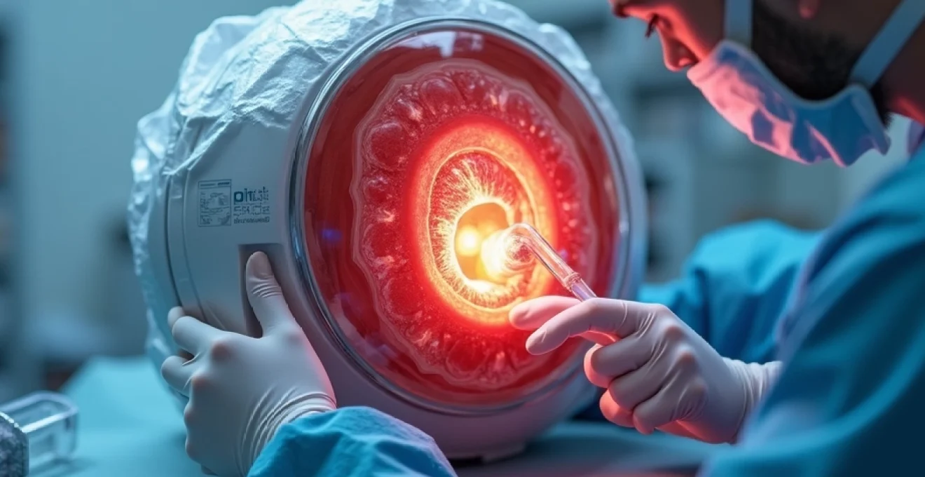
Ultrasonic phacoemulsification technique using alcon centurion vision system
Contemporary phacoemulsification employs sophisticated ultrasonic technology to break apart the cataractous lens whilst preserving the delicate surrounding eye structures. The Alcon Centurion Vision System represents the pinnacle of this technology, utilising active fluidics management to maintain stable anterior chamber pressure throughout the procedure. This system delivers precise ultrasonic energy through a titanium tip oscillating at 40,000 Hz, enabling surgeons to fragment even the densest cataracts with minimal trauma to adjacent tissues.
The Centurion platform incorporates intelligent software algorithms that automatically adjust aspiration flow rates and vacuum levels based on real-time surgical conditions. Surgeons can customise parameters including longitudinal ultrasound power, torsional amplitude, and aspiration settings to match individual patient anatomy and cataract density. This personalised approach significantly reduces the risk of complications such as posterior capsule rupture or corneal endothelial damage.
Monofocal versus multifocal IOL selection: alcon AcrySof and johnson & johnson tecnis
Intraocular lens selection represents a crucial decision point in cataract surgery, directly impacting patient satisfaction and visual outcomes. Monofocal IOLs provide excellent distance vision but typically require reading glasses for near tasks, whilst multifocal designs offer vision at multiple distances with potential trade-offs in contrast sensitivity and night vision quality. The Alcon AcrySof platform includes both monofocal and multifocal options, with the AcrySof IQ PanOptix representing their latest trifocal design.
Johnson & Johnson’s Tecnis IOL family offers advanced aspheric optics designed to reduce spherical aberration and improve contrast sensitivity. The Tecnis Symfony extended depth-of-focus lens provides continuous vision from distance to intermediate range whilst minimising the visual disturbances sometimes associated with traditional multifocal designs. Recent clinical data suggests patient satisfaction rates exceeding 90% when appropriate IOL selection criteria are followed.
Femtosecond Laser-Assisted cataract surgery (FLACS) with LenSx platform
Femtosecond laser technology has introduced unprecedented precision to cataract surgery, enabling surgeons to create perfectly circular capsulotomies and precise corneal incisions with micron-level accuracy. The LenSx laser platform utilises infrared light pulses lasting just 10^-15 seconds to create precise tissue separations without generating heat or causing collateral damage. This technology particularly benefits complex cases involving dense cataracts or challenging anatomy.
FLACS procedures demonstrate reduced effective phacoemulsification time and decreased ultrasonic energy requirements compared to traditional manual techniques. Studies indicate that laser-assisted capsulotomies show superior circularity and centration compared to manual capsulorhexis, potentially improving IOL positioning and long-term stability. The enhanced precision offered by FLACS makes it particularly valuable for premium IOL implantation where optimal lens positioning is critical for achieving desired refractive outcomes.
Post-operative endophthalmitis prevention protocols and antibiotic prophylaxis
Endophthalmitis, though rare with an incidence of approximately 0.1%, represents the most serious potential complication following cataract surgery. Contemporary prevention protocols emphasise multi-modal approaches including pre-operative antisepsis, intracameral antibiotic injection, and careful attention to surgical sterility. Povidone-iodine 5% remains the gold standard for pre-operative conjunctival preparation, whilst intracameral cefuroxime or moxifloxacin injection at surgery conclusion has demonstrated significant efficacy in reducing infection rates.
Modern endophthalmitis prevention protocols have reduced infection rates by over 80% compared to historical controls, making cataract surgery one of the safest procedures in contemporary medicine.
Laparoscopic cholecystectomy: minimally invasive gallbladder removal
Laparoscopic cholecystectomy has become the definitive treatment for symptomatic gallstone disease and acute cholecystitis, representing one of the most commonly performed abdominal procedures worldwide. This minimally invasive approach offers significant advantages over traditional open surgery, including reduced post-operative pain, shorter hospital stays, faster recovery times, and superior cosmetic outcomes. The procedure typically involves four small incisions rather than a single large abdominal opening, fundamentally changing the patient experience and recovery trajectory.
Current data indicates that over 750,000 cholecystectomies are performed annually in the United States alone, with laparoscopic techniques accounting for approximately 95% of all cases. The conversion rate to open surgery has decreased significantly with improved surgical techniques and enhanced imaging technology, now averaging less than 5% in most centres. Patient satisfaction rates consistently exceed 90%, with most individuals returning to normal activities within one to two weeks following surgery.
Four-port laparoscopic technique with CO2 insufflation parameters
The standard four-port laparoscopic cholecystectomy technique involves strategically placed trocars to optimise surgical access whilst minimising patient discomfort. The 12mm umbilical port accommodates the laparoscope and specimen extraction, whilst three 5mm ports provide working channels for grasping and dissection instruments. Pneumoperitoneum creation using CO2 insufflation at 12-15 mmHg provides adequate working space whilst maintaining patient safety through rapid gas absorption and elimination.
Optimal port placement follows anatomical landmarks to ensure proper triangulation and avoid injury to underlying structures. The epigastric port, positioned in the midline below the xiphoid process, serves as the primary working port for dissection. Lateral subcostal ports at the midclavicular and anterior axillary lines provide traction and counter-traction capabilities essential for safe tissue dissection. Modern insufflation systems incorporate pressure monitoring and automatic flow regulation to maintain stable pneumoperitoneum throughout the procedure.
Critical view of safety identification and calot’s triangle dissection
The Critical View of Safety represents the fundamental principle ensuring safe laparoscopic cholecystectomy performance, requiring clear identification of specific anatomical landmarks before proceeding with clip application or division of any structures. This safety criterion mandates visualisation of only two structures entering the gallbladder: the cystic artery and cystic duct, with clear identification of the common hepatic duct and proper hepatic artery.
Calot’s triangle dissection requires meticulous technique to avoid injury to the common bile duct or aberrant arterial anatomy. Systematic dissection proceeds from lateral to medial , carefully clearing inflammatory tissue and adhesions whilst preserving critical structures. Studies demonstrate that strict adherence to Critical View of Safety principles reduces bile duct injury rates to less than 0.3%, significantly lower than historical rates associated with traditional techniques.
Single-incision laparoscopic surgery (SILS) using SPIDER surgical system
Single-incision laparoscopic surgery represents the evolution towards even less invasive surgical approaches, utilising a single umbilical incision for all instrumentation. The SPIDER (Single Port Instrument Delivery Extended Reach) system provides flexible instruments capable of triangulation through a single port, maintaining surgical precision whilst minimising abdominal wall trauma. This technology addresses previous limitations of single-port surgery, including instrument collision and reduced ergonomics.
SILS cholecystectomy offers potential advantages including reduced post-operative pain, improved cosmetic outcomes, and faster recovery compared to traditional multi-port techniques. Patient selection remains crucial for optimal outcomes, with ideal candidates including those with uncomplicated gallstone disease and minimal inflammatory changes. Recent meta-analyses suggest comparable safety profiles to conventional laparoscopy, though longer operative times and steeper learning curves remain considerations.
Intraoperative cholangiography protocol and bile duct injury management
Intraoperative cholangiography serves both diagnostic and therapeutic purposes during laparoscopic cholecystectomy, providing real-time assessment of biliary anatomy and identifying potential injuries or retained stones. Modern fluoroscopic imaging allows dynamic visualisation of contrast flow through the biliary system, enabling immediate recognition of anatomical variants or complications. Contrast injection protocols typically utilise 50% diluted contrast medium to optimise visualisation whilst minimising viscosity-related flow issues.
Systematic use of intraoperative cholangiography can reduce major bile duct injury rates and improve outcomes when complications do occur through early recognition and immediate management.
Arthroscopic knee surgery: meniscal repair and ACL reconstruction
Arthroscopic knee surgery has revolutionised orthopaedic practice, transforming complex joint procedures into minimally invasive operations with dramatically improved patient outcomes. This advanced surgical technique utilises small incisions and specialised instruments to address various knee pathologies, including meniscal tears, ligament injuries, and cartilage damage. The arthroscopic approach offers significant advantages over traditional open surgery, including reduced tissue trauma, decreased post-operative pain, faster rehabilitation, and superior cosmetic results.
Contemporary arthroscopic procedures benefit from high-definition imaging systems and advanced instrumentation that enable surgeons to perform complex repairs with exceptional precision. Modern arthroscopes provide 4K resolution imagery with enhanced colour definition and depth perception, allowing detailed assessment of intra-articular structures. Annual statistics indicate that over 2 million arthroscopic knee procedures are performed globally, with success rates consistently exceeding 90% for most indications.
Anteromedial and anterolateral portal placement techniques
Proper portal placement represents the foundation of successful arthroscopic knee surgery, directly impacting visualisation quality and instrument access to all joint compartments. The anteromedial portal typically serves as the primary viewing portal, positioned approximately 1cm medial to the patellar tendon and 1cm above the joint line. This location provides optimal visualisation of the lateral compartment whilst minimising risk to the infrapatellar branch of the saphenous nerve.
The anterolateral portal functions as the primary working portal, positioned 1cm lateral to the patellar tendon at the joint line level. Precise portal placement ensures triangulation between instruments whilst avoiding neurovascular structures and maintaining adequate working space. Advanced techniques may incorporate additional portals for complex procedures, including posteromedial or posterolateral approaches for addressing posterior horn pathology or performing comprehensive meniscal repairs.
All-inside ACL reconstruction using quadriceps tendon autograft
All-inside anterior cruciate ligament reconstruction represents a significant advancement in knee ligament surgery, utilising adjustable-loop cortical fixation devices that eliminate the need for femoral cross-pin or interference screw fixation. This technique reduces surgical trauma whilst providing biomechanically superior fixation strength compared to traditional methods. Quadriceps tendon autografts have gained popularity due to their favourable biomechanical properties and reduced harvest site morbidity compared to bone-patellar tendon-bone grafts.
The quadriceps tendon provides adequate graft length for all-inside techniques whilst maintaining excellent tensile strength characteristics. Graft preparation protocols typically involve harvesting a 10-12mm wide central portion of the quadriceps tendon, preserving the surrounding muscle fibres and minimising functional deficits. Clinical studies demonstrate return-to-sport rates exceeding 85% following all-inside ACL reconstruction, with patient satisfaction scores consistently high due to reduced post-operative pain and faster rehabilitation progression.
Meniscal root repair with suture anchor fixation methods
Meniscal root tears represent complex injuries requiring specialised repair techniques to restore normal knee biomechanics and prevent progressive joint degeneration. These tears occur at the attachment points where the meniscus anchors to the tibial plateau, disrupting the circumferential hoop stress distribution essential for joint protection. Root repair procedures have evolved to incorporate suture anchor fixation systems that provide reliable anatomical restoration of the meniscal attachment.
Modern suture anchor systems utilise biocompatible materials and advanced thread designs to optimise healing potential whilst providing immediate mechanical stability. The repair process involves anatomical reduction of the torn meniscal root to its original footprint, followed by secure fixation using multiple suture anchor points. Long-term studies indicate that successful meniscal root repair can prevent or significantly delay the progression of osteoarthritis, making this procedure increasingly valuable for preserving joint health.
Post-arthroscopic rehabilitation protocols and Return-to-Sport criteria
Contemporary rehabilitation following arthroscopic knee surgery emphasises evidence-based protocols tailored to specific procedures and individual patient goals. Early mobilisation principles promote faster recovery whilst protecting healing tissues through graduated loading progressions. Protocol development considers factors including tissue healing timelines, biomechanical demands of sporting activities, and psychological readiness for return to full activity levels.
Successful return to sport following arthroscopic knee surgery depends not only on surgical technique but equally on adherence to comprehensive rehabilitation protocols that address strength, proprioception, and sport-specific movement patterns.
Coronary artery bypass grafting (CABG): On-Pump and Off-Pump techniques
Coronary artery bypass grafting remains the gold standard treatment for complex coronary artery disease, particularly in patients with multi-vessel disease, left main coronary artery stenosis, or diabetes mellitus. This sophisticated cardiac procedure involves creating alternative pathways around blocked coronary arteries using harvested vessels from other parts of the patient’s body. CABG procedures demonstrate exceptional long-term durability, with many grafts remaining patent for decades following surgery.
Modern cardiac surgery centres perform thousands of CABG procedures annually, with mortality rates consistently below 2% in most developed healthcare systems. The choice between on-pump and off-pump techniques depends on patient anatomy, surgeon expertise, and specific clinical circumstances. Recent advances in surgical techniques, perfusion technology, and myocardial protection strategies have further improved outcomes whilst expanding the procedure’s applicability to higher-risk patient populations.
On-pump CABG utilises cardiopulmonary bypass to maintain circulation whilst the heart is temporarily stopped, providing optimal surgical conditions for precise anastomotic construction. This traditional approach offers excellent visualisation and facilitates complex multi-vessel revascularisation procedures. Modern perfusion systems incorporate biocompatible circuits, advanced oxygenators, and sophisticated monitoring systems that minimise blood trauma whilst maintaining optimal organ perfusion throughout the procedure.
Off-pump coronary artery bypass represents an alternative approach that avoids cardiopulmonary bypass whilst maintaining myocardial perfusion during anastomotic construction. This technique utilises mechanical stabilisation systems and specialised retraction devices to immobilise specific coronary artery segments whilst the remainder of the heart continues beating. Studies suggest potential advantages including reduced inflammatory response, decreased bleeding complications, and faster post-operative recovery in selected patient populations.
Appendectomy procedures: open McBurney versus laparoscopic approaches
Appendectomy stands as one of the most commonly performed emergency surgical procedures worldwide, with over 300
,000 procedures performed annually in the United States alone. This fundamental surgical intervention addresses acute appendicitis, one of the most common abdominal emergencies requiring immediate surgical attention. The evolution from open McBurney incision techniques to minimally invasive laparoscopic approaches has significantly transformed patient outcomes, recovery times, and overall surgical experience.
Modern appendectomy techniques demonstrate remarkably low complication rates, with mortality approaching zero in uncomplicated cases. The choice between open and laparoscopic approaches depends on factors including patient presentation, surgeon expertise, available resources, and specific clinical circumstances. Both techniques offer excellent outcomes when performed appropriately, though laparoscopic methods increasingly dominate contemporary practice due to their minimally invasive nature.
The traditional McBurney incision, named after surgeon Charles McBurney, involves a focused right lower quadrant approach through a small oblique incision positioned over McBurney’s point. This technique provides direct access to the appendix whilst minimising tissue trauma and maintaining excellent cosmetic results. Incision placement follows anatomical landmarks approximately one-third the distance from the anterior superior iliac spine to the umbilicus, allowing optimal visualisation of the appendiceal region.
Laparoscopic appendectomy utilises three small port incisions to remove the inflamed appendix using advanced visualisation and instrumentation systems. This approach offers significant advantages including reduced post-operative pain, faster recovery, shorter hospital stays, and superior diagnostic capabilities for atypical presentations. Studies consistently demonstrate earlier return to normal activities and higher patient satisfaction scores compared to open techniques, making laparoscopy the preferred approach in suitable candidates.
Contemporary laparoscopic appendectomy demonstrates conversion rates to open surgery of less than 5% in most centres, with complication rates significantly lower than historical open surgery series.
Total hip replacement: anterior and posterior surgical approaches
Total hip replacement represents one of the most successful surgical interventions in modern orthopaedic practice, consistently ranking among the most effective procedures for improving quality of life and functional outcomes. This comprehensive joint reconstruction addresses severe arthritis, fractures, and degenerative conditions that cause debilitating pain and mobility limitations. With over 450,000 hip replacement procedures performed annually in the United States, this surgery has transformed millions of lives through restoration of pain-free mobility and independence.
The longevity of modern hip implants exceeds 20 years in the majority of patients, with some contemporary designs demonstrating even greater durability. Advanced bearing surfaces and improved implant designs have virtually eliminated historical concerns regarding wear and loosening, making hip replacement a reliable long-term solution for joint disease. Patient satisfaction rates consistently exceed 95%, with most individuals experiencing dramatic pain relief and functional improvement within weeks of surgery.
The anterior approach to total hip replacement utilises the natural interval between muscle groups to access the hip joint without cutting major muscles or tendons. This technique preserves the integrity of the posterior capsule and critical soft tissue structures, potentially reducing dislocation risk and accelerating recovery. Anterior approach advocates emphasise benefits including reduced post-operative restrictions, faster rehabilitation, and improved early functional outcomes compared to traditional posterior approaches.
Specialised anterior approach tables facilitate optimal patient positioning whilst providing fluoroscopic imaging capabilities for enhanced component positioning accuracy. The technique requires extensive surgical experience due to the limited surgical exposure and proximity to neurovascular structures. Studies suggest that anterior approach patients may experience faster early recovery and reduced length of stay, though long-term outcomes appear similar to posterior approaches when performed by experienced surgeons.
The posterior approach remains the most widely utilised technique for total hip replacement, offering excellent surgical exposure and versatility for complex cases. This traditional method provides optimal visualisation of the acetabulum and proximal femur whilst accommodating various implant designs and surgical challenges. Posterior approach modifications including enhanced soft tissue repair and capsular reconstruction have significantly reduced historical dislocation concerns.
Contemporary posterior hip replacement incorporates advanced surgical techniques including minimally invasive incisions, tissue-sparing approaches, and enhanced recovery protocols. The technique offers advantages for revision surgeries, complex deformities, and challenging anatomical situations where maximum surgical exposure proves beneficial. Surgeons can adapt the approach based on patient anatomy and specific requirements, making it versatile for diverse clinical scenarios.
Modern total hip replacement surgery benefits from advanced imaging technologies, computer-assisted navigation systems, and robotically-assisted surgical platforms that enhance precision and reproducibility. These technological innovations enable optimal component positioning, improved limb length restoration, and reduced risk of complications. Patient-specific instrumentation and pre-operative planning utilising three-dimensional imaging further optimise surgical outcomes whilst reducing operative time and complexity.
Recovery following total hip replacement has been revolutionised through enhanced recovery after surgery (ERAS) protocols that emphasise multimodal pain management, early mobilisation, and patient education. Most patients now ambulate within hours of surgery and achieve functional independence within days rather than weeks. Contemporary rehabilitation programmes focus on strength restoration, gait training, and activity progression to ensure optimal long-term outcomes and patient satisfaction.